Understanding the Structural Basis for Substrate and Inhibitor Recognition in Eukaryotic Gh11 Xylanases.
Vardakou, M., Dumon, C., Murray, J.W., Christakopoulos, P., Weiner, D.P., Juge, N., Lewis, R.J., Gilbert, H.J., Flint, J.E.(2008) J Mol Biol 375: 1293
- PubMed: 18078955
- DOI: https://doi.org/10.1016/j.jmb.2007.11.007
- Primary Citation of Related Structures:
2C1F, 2VG9, 2VGD - PubMed Abstract:
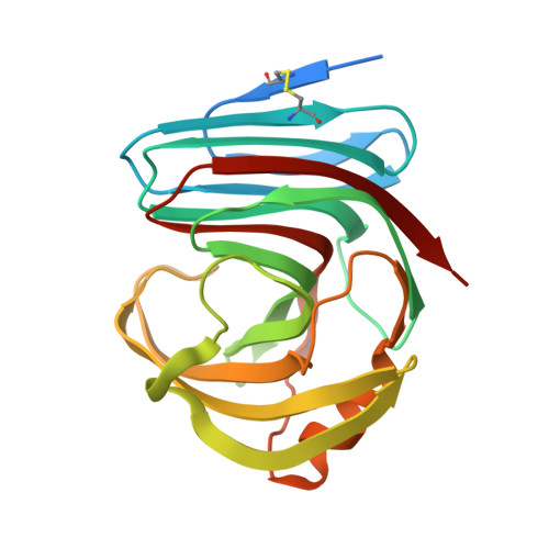
Endo-beta1,4-xylanases (xylanases) hydrolyse the beta1,4 glycosidic bonds in the backbone of xylan. Although xylanases from glycoside hydrolase family 11 (GH11) have been extensively studied, several issues remain unresolved. Thus, the mechanism by which these enzymes hydrolyse decorated xylans is unclear and the structural basis for the variation in catalytic activity within this family is unknown. Furthermore, the mechanism for the differences in the inhibition of fungal GH11 enzymes by the wheat protein XIP-I remains opaque. To address these issues we report the crystal structure and biochemical properties of the Neocallimastix patriciarum xylanase NpXyn11A, which displays unusually high catalytic activity and is one of the few fungal GH11 proteins not inhibited by XIP-I. Although the structure of NpXyn11A could not be determined in complex with substrates, we have been able to investigate how GH11 enzymes hydrolyse decorated substrates by solving the crystal structure of a second GH11 xylanase, EnXyn11A (encoded by an environmental DNA sample), bound to ferulic acid-1,5-arabinofuranose-alpha1,3-xylotriose (FAX(3)). The crystal structure of the EnXyn11A-FAX(3) complex shows that solvent exposure of the backbone xylose O2 and O3 groups at subsites -3 and +2 allow accommodation of alpha1,2-linked 4-methyl-D-glucuronic acid and L-arabinofuranose side chains. Furthermore, the ferulated arabinofuranose side chain makes hydrogen bonds and hydrophobic interactions at the +2 subsite, indicating that the decoration may represent a specificity determinant at this aglycone subsite. The structure of NpXyn11A reveals potential -3 and +3 subsites that are kinetically significant. The extended substrate-binding cleft of NpXyn11A, compared to other GH11 xylanases, may explain why the Neocallimastix enzyme displays unusually high catalytic activity. Finally, the crystal structure of NpXyn11A shows that the resistance of the enzyme to XIP-I is not due solely to insertions in the loop connecting beta strands 11 and 12, as suggested previously, but is highly complex.
Organizational Affiliation:
Institute for Cell and Molecular Biosciences, Newcastle University, The Medical School, Framlington Place, Newcastle upon Tyne NE2 4HH, UK.