Divergence of Cofactor Recognition Across Evolution: Coenzyme a Binding in a Prokaryotic Arylamine N-Acetyltransferase.
Fullam, E., Westwood, I.M., Anderton, M.C., Lowe, E.D., Sim, E., Noble, M.E.M.(2008) J Mol Biol 375: 178
- PubMed: 18005984
- DOI: https://doi.org/10.1016/j.jmb.2007.10.019
- Primary Citation of Related Structures:
2VFB, 2VFC - PubMed Abstract:
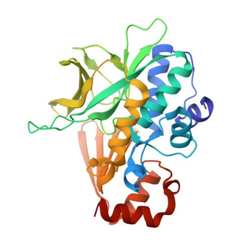
Arylamine N-acetyltransferase (NAT) enzymes are widespread in nature. They serve to acetylate xenobiotics and/or endogenous substrates using acetyl coenzyme A (CoA) as a cofactor. Conservation of the architecture of the NAT enzyme family from mammals to bacteria has been demonstrated by a series of prokaryotic NAT structures, together with the recently reported structure of human NAT1. We report here the cloning, purification, kinetic characterisation and crystallographic structure determination of NAT from Mycobacterium marinum, a close relative of the pathogenic Mycobacterium tuberculosis. We have also determined the structure of M. marinum NAT in complex with CoA, shedding the first light on cofactor recognition in prokaryotic NATs. Surprisingly, the principal CoA recognition site in M. marinum NAT is located some 30 A from the site of CoA recognition in the recently deposited structure of human NAT2 bound to CoA. The structure explains the Ping-Pong Bi-Bi reaction mechanism of NAT enzymes and suggests mechanisms by which the acetylated enzyme intermediate may be protected. Recognition of CoA in a much wider groove in prokaryotic NATs suggests that this subfamily may accommodate larger substrates than is the case for human NATs and may assist in the identification of potential endogenous substrates. It also suggests the cofactor-binding site as a unique subsite to target in drug design directed against NAT in mycobacteria.
Organizational Affiliation:
Department of Pharmacology, University of Oxford, Mansfield Road, Oxford OX1 3QT, UK.