2VF2

X-ray crystal structure of HsaD from Mycobacterium tuberculosis


Experimental Data Snapshot

  • Method: X-RAY DIFFRACTION
  • Resolution: 2.35 Å
  • R-Value Free: 0.233 
  • R-Value Work: 0.212 

wwPDB Validation   3D Report Full Report


This is version 1.3 of the entry. See complete history


Literature

Structure of Hsad, a Steroid-Degrading Hydrolase, from Mycobacterium Tuberculosis.

Lack, N.Lowe, E.D.Liu, J.Eltis, L.D.Noble, M.E.M.Sim, E.Westwood, I.M.

(2008) Acta Crystallogr Sect F Struct Biol Cryst Commun 64: 2

  • DOI: https://doi.org/10.1107/S1744309107065931
  • Primary Citation of Related Structures:  
    2VF2

  • PubMed Abstract: 

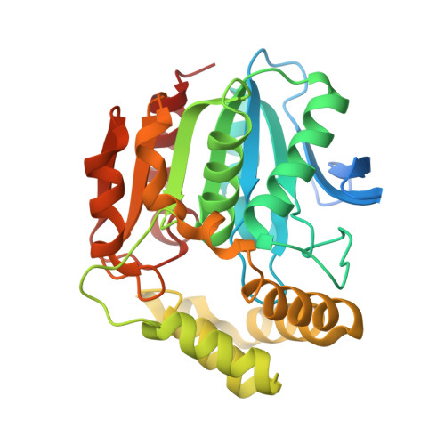
    Tuberculosis is a major cause of death worldwide. Understanding of the pathogenicity of Mycobacterium tuberculosis has been advanced by gene analysis and has led to the identification of genes that are important for intracellular survival in macrophages. One of these genes encodes HsaD, a meta-cleavage product (MCP) hydrolase that catalyzes the hydrolytic cleavage of a carbon-carbon bond in cholesterol metabolism. This paper describes the production of HsaD as a recombinant protein and, following crystallization, the determination of its three-dimensional structure to 2.35 A resolution by X-ray crystallography at the Diamond Light Source in Oxfordshire, England. To the authors' knowledge, this study constitutes the first report of a structure determined at the new synchrotron facility. The volume of the active-site cleft of the HsaD enzyme is more than double the corresponding active-site volumes of related MCP hydrolases involved in the catabolism of aromatic compounds, consistent with the specificity of HsaD for steroids such as cholesterol. Knowledge of the structure of the enzyme facilitates the design of inhibitors.


  • Organizational Affiliation

    Department of Pharmacology, University of Oxford, Mansfield Road, Oxford OX1 3QT, England.


Macromolecules
Find similar proteins by:  (by identity cutoff)  |  3D Structure
Entity ID: 1
MoleculeChains Sequence LengthOrganismDetailsImage
2-HYDROXY-6-OXO-6-PHENYLHEXA-2,4-DIENOATE HYDROLASE BPHD
A, B
311Mycobacterium tuberculosis H37RvMutation(s): 0 
EC: 3.7.1.8
UniProt
Find proteins for P9WNH5 (Mycobacterium tuberculosis (strain ATCC 25618 / H37Rv))
Explore P9WNH5 
Go to UniProtKB:  P9WNH5
Entity Groups  
Sequence Clusters30% Identity50% Identity70% Identity90% Identity95% Identity100% Identity
UniProt GroupP9WNH5
Sequence Annotations
Expand
  • Reference Sequence
Experimental Data & Validation

Experimental Data

  • Method: X-RAY DIFFRACTION
  • Resolution: 2.35 Å
  • R-Value Free: 0.233 
  • R-Value Work: 0.212 
  • Space Group: I 21 21 21
Unit Cell:
Length ( Å )Angle ( ˚ )
a = 82.36α = 90
b = 82.46β = 90
c = 194.67γ = 90
Software Package:
Software NamePurpose
PHENIXrefinement
MOSFLMdata reduction
SCALAdata scaling
PHASERphasing
PHENIXrefinement

Structure Validation

View Full Validation Report


Entry History 

Deposition Data

Revision History  (Full details and data files)

  • Version 1.0: 2007-11-06
    Type: Initial release
  • Version 1.1: 2011-05-08
    Changes: Version format compliance
  • Version 1.2: 2011-07-13
    Changes: Version format compliance
  • Version 1.3: 2023-12-13
    Changes: Data collection, Database references, Other, Refinement description