2VES

Crystal Structure of LpxC from Pseudomonas aeruginosa complexed with the potent BB-78485 inhibitor


Experimental Data Snapshot

  • Method: X-RAY DIFFRACTION
  • Resolution: 1.90 Å
  • R-Value Free: 0.221 
  • R-Value Work: 0.177 
  • R-Value Observed: 0.179 

wwPDB Validation   3D Report Full Report


Ligand Structure Quality Assessment 


This is version 1.5 of the entry. See complete history


Literature

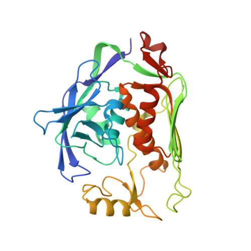
Crystal structure of LpxC from Pseudomonas aeruginosa complexed with the potent BB-78485 inhibitor.

Mochalkin, I.Knafels, J.D.Lightle, S.

(2008) Protein Sci 17: 450-457

  • DOI: https://doi.org/10.1110/ps.073324108
  • Primary Citation of Related Structures:  
    2VES

  • PubMed Abstract: 

    The cell wall in Gram-negative bacteria is surrounded by an outer membrane comprised of charged lipopolysaccharide (LPS) molecules that prevent entry of hydrophobic agents into the cell and protect the bacterium from many antibiotics. The hydrophobic anchor of LPS is lipid A, the biosynthesis of which is essential for bacterial growth and viability. UDP-3-O-(R-3-hydroxymyristoyl)-N-acetylglucosamine deacetylase (LpxC) is an essential zinc-dependant enzyme that catalyzes the conversion of UDP-3-O-(R-3-hydroxymyristoyl)-N-acetylglucosamine to UDP-3-O-(R-3-hydroxymyristoyl)glucosamine and acetate in the biosynthesis of lipid A, and for this reason, LpxC is an attractive target for antibacterial drug discovery. Here we disclose a 1.9 A resolution crystal structure of LpxC from Pseudomonas aeruginosa (paLpxC) in a complex with the potent BB-78485 inhibitor. To our knowledge, this is the first crystal structure of LpxC with a small-molecule inhibitor that shows antibacterial activity against a wide range of Gram-negative pathogens. Accordingly, this structure can provide important information for lead optimization and rational design of the effective small-molecule LpxC inhibitors for successful treatment of Gram-negative infections.


  • Organizational Affiliation

    Pfizer, Inc., Groton, Connecticut 06340, USA. Igor.Mochalkin@pfizer.com


Macromolecules
Find similar proteins by:  (by identity cutoff)  |  3D Structure
Entity ID: 1
MoleculeChains Sequence LengthOrganismDetailsImage
UDP-3-O-acyl-N-acetylglucosamine deacetylase
A, B, C
299Pseudomonas aeruginosa PAO1Mutation(s): 0 
Gene Names: lpxCenvAPA4406
EC: 3.5.1.108
UniProt
Find proteins for P47205 (Pseudomonas aeruginosa (strain ATCC 15692 / DSM 22644 / CIP 104116 / JCM 14847 / LMG 12228 / 1C / PRS 101 / PAO1))
Explore P47205 
Go to UniProtKB:  P47205
Entity Groups  
Sequence Clusters30% Identity50% Identity70% Identity90% Identity95% Identity100% Identity
UniProt GroupP47205
Sequence Annotations
Expand
  • Reference Sequence
Small Molecules
Ligands 3 Unique
IDChains Name / Formula / InChI Key2D Diagram3D Interactions
GVR
Query on GVR

Download Ideal Coordinates CCD File 
E [auth A],
H [auth B],
L [auth C]
(2R)-N-hydroxy-3-naphthalen-2-yl-2-[(naphthalen-2-ylsulfonyl)amino]propanamide
C23 H20 N2 O4 S
MMOUXLMPQFMDRD-JOCHJYFZSA-N
SO4
Query on SO4

Download Ideal Coordinates CCD File 
J [auth B],
O [auth C]
SULFATE ION
O4 S
QAOWNCQODCNURD-UHFFFAOYSA-L
ZN
Query on ZN

Download Ideal Coordinates CCD File 
D [auth A]
F [auth A]
G [auth B]
I [auth B]
K [auth C]
D [auth A],
F [auth A],
G [auth B],
I [auth B],
K [auth C],
M [auth C],
N [auth C]
ZINC ION
Zn
PTFCDOFLOPIGGS-UHFFFAOYSA-N
Binding Affinity Annotations 
IDSourceBinding Affinity
GVR PDBBind:  2VES Ki: 20 (nM) from 1 assay(s)
Experimental Data & Validation

Experimental Data

  • Method: X-RAY DIFFRACTION
  • Resolution: 1.90 Å
  • R-Value Free: 0.221 
  • R-Value Work: 0.177 
  • R-Value Observed: 0.179 
  • Space Group: P 21 21 21
Unit Cell:
Length ( Å )Angle ( ˚ )
a = 90.542α = 90
b = 103.331β = 90
c = 105.284γ = 90
Software Package:
Software NamePurpose
REFMACrefinement
HKL-2000data reduction
HKL-2000data scaling
MOLREPphasing

Structure Validation

View Full Validation Report


Ligand Structure Quality Assessment 


Entry History 

Deposition Data

Revision History  (Full details and data files)

  • Version 1.0: 2008-01-15
    Type: Initial release
  • Version 1.1: 2011-05-08
    Changes: Version format compliance
  • Version 1.2: 2011-07-13
    Changes: Version format compliance
  • Version 1.3: 2018-09-19
    Changes: Data collection, Database references, Source and taxonomy, Structure summary
  • Version 1.4: 2019-05-08
    Changes: Data collection, Experimental preparation
  • Version 1.5: 2023-12-13
    Changes: Data collection, Database references, Derived calculations, Other, Refinement description