2VEP

Crystal Structure Of The Full Length Bifunctional Enzyme Pria


Experimental Data Snapshot

  • Method: X-RAY DIFFRACTION
  • Resolution: 1.80 Å
  • R-Value Free: 0.224 
  • R-Value Work: 0.180 
  • R-Value Observed: 0.181 

wwPDB Validation   3D Report Full Report


This is version 1.2 of the entry. See complete history


Literature

The Structure/Function Relationship of a Dual Substrate (Betaalpha)(8)-Isomerase

Wright, H.Noda-Garcia, L.Ochoa-Leyva, A.Hodgson, D.A.Fulop, V.Barona-Gomez, F.

(2008) Biochem Biophys Res Commun 365: 16

  • DOI: https://doi.org/10.1016/j.bbrc.2007.10.101
  • Primary Citation of Related Structures:  
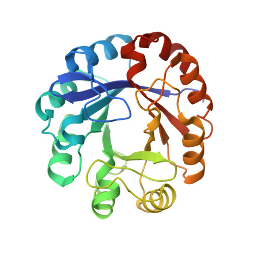
    2VEP

  • PubMed Abstract: 

    Two structures of phosphoribosyl isomerase A (PriA) from Streptomyces coelicolor, involved in both histidine and tryptophan biosynthesis, were solved at 1.8A resolution. A closed conformer was obtained, which represents the first complete structure of PriA, revealing hitherto unnoticed molecular interactions and the occurrence of conformational changes. Inspection of these conformers, including ligand-docking simulations, allowed identification of residues involved in substrate recognition, chemical catalysis and conformational changes. These predictions were validated by mutagenesis and functional analysis. Arg19 and Ser81 were shown to play critical roles within the carboxyl and amino phosphate-binding sites, respectively; the catalytic residues Asp11 and Asp130 are responsible for both activities; and Thr166 and Asp171, which make an unusual contact, are likely to elicit the conformational changes needed for adopting the active site architectures. This represents the first report of the structure/function relationship of this (betaalpha)8-isomerase.


  • Organizational Affiliation

    Department of Biological Sciences, University of Warwick, Gibbet Hill Road, Coventry CV4 7AL, UK.


Macromolecules
Find similar proteins by:  (by identity cutoff)  |  3D Structure
Entity ID: 1
MoleculeChains Sequence LengthOrganismDetailsImage
PHOSPHORIBOSYL ISOMERASE A244Streptomyces coelicolorMutation(s): 0 
EC: 5.3.1.16 (PDB Primary Data), 5.3.1.24 (PDB Primary Data)
UniProt
Find proteins for P16250 (Streptomyces coelicolor (strain ATCC BAA-471 / A3(2) / M145))
Explore P16250 
Go to UniProtKB:  P16250
Entity Groups  
Sequence Clusters30% Identity50% Identity70% Identity90% Identity95% Identity100% Identity
UniProt GroupP16250
Sequence Annotations
Expand
  • Reference Sequence
Experimental Data & Validation

Experimental Data

  • Method: X-RAY DIFFRACTION
  • Resolution: 1.80 Å
  • R-Value Free: 0.224 
  • R-Value Work: 0.180 
  • R-Value Observed: 0.181 
  • Space Group: P 31 2 1
Unit Cell:
Length ( Å )Angle ( ˚ )
a = 65.14α = 90
b = 65.14β = 90
c = 104.67γ = 120
Software Package:
Software NamePurpose
REFMACrefinement
DENZOdata reduction
SCALEPACKdata scaling
SOLVEphasing

Structure Validation

View Full Validation Report


Entry History 

Deposition Data

Revision History  (Full details and data files)

  • Version 1.0: 2007-11-20
    Type: Initial release
  • Version 1.1: 2011-07-13
    Changes: Advisory, Version format compliance
  • Version 1.2: 2018-01-24
    Changes: Source and taxonomy