2VDV

Structure of trm8, m7G methylation enzyme


Experimental Data Snapshot

  • Method: X-RAY DIFFRACTION
  • Resolution: 2.30 Å
  • R-Value Free: 0.265 
  • R-Value Work: 0.233 

wwPDB Validation   3D Report Full Report


Ligand Structure Quality Assessment 


This is version 1.2 of the entry. See complete history


Literature

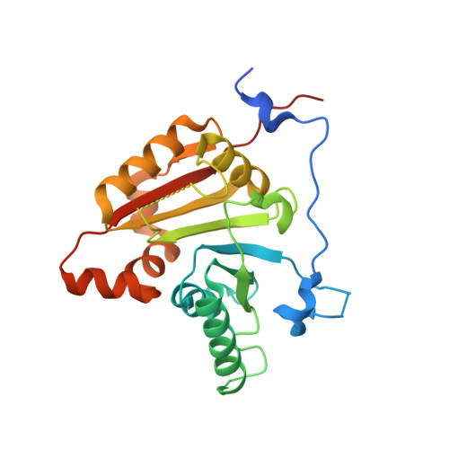
Structure of the Yeast tRNA M7G Methylation Complex.

Leulliot, N.Chaillet, M.Durand, D.Ulryck, N.Blondeau, K.Van Tilbeurgh, H.

(2008) Structure 16: 52

  • DOI: https://doi.org/10.1016/j.str.2007.10.025
  • Primary Citation of Related Structures:  
    2VDU, 2VDV

  • PubMed Abstract: 

    Loss of N7-methylguanosine (m7G) modification is involved in the recently discovered rapid tRNA degradation pathway. In yeast, this modification is catalyzed by the heterodimeric complex composed of a catalytic subunit Trm8 and a noncatalytic subunit Trm82. We have solved the crystal structure of Trm8 alone and in complex with Trm82. Trm8 undergoes subtle conformational changes upon Trm82 binding which explains the requirement of Trm82 for activity. Cocrystallization with the S-adenosyl-methionine methyl donor defines the putative catalytic site and a guanine binding pocket. Small-angle X-ray scattering in solution of the Trm8-Trm82 heterodimer in complex with tRNA(Phe) has enabled us to propose a low-resolution structure of the ternary complex which defines the tRNA binding mode of Trm8-Trm82 and the structural elements contributing to specificity.


  • Organizational Affiliation

    Institut de Biochimie et de Biophysique Moléculaire et Cellulaire, UMR8619, Bât 430, Université de Paris-Sud, IFR115, Orsay Cedex, France. nicolas.leulliot@u-psud.fr


Macromolecules
Find similar proteins by:  (by identity cutoff)  |  3D Structure
Entity ID: 1
MoleculeChains Sequence LengthOrganismDetailsImage
TRNA (GUANINE-N(7)-)-METHYLTRANSFERASEA [auth E],
B [auth F]
246Saccharomyces cerevisiaeMutation(s): 0 
EC: 2.1.1.33
UniProt
Find proteins for Q12009 (Saccharomyces cerevisiae (strain ATCC 204508 / S288c))
Explore Q12009 
Go to UniProtKB:  Q12009
Entity Groups  
Sequence Clusters30% Identity50% Identity70% Identity90% Identity95% Identity100% Identity
UniProt GroupQ12009
Sequence Annotations
Expand
  • Reference Sequence
Small Molecules
Experimental Data & Validation

Experimental Data

  • Method: X-RAY DIFFRACTION
  • Resolution: 2.30 Å
  • R-Value Free: 0.265 
  • R-Value Work: 0.233 
  • Space Group: C 1 2 1
Unit Cell:
Length ( Å )Angle ( ˚ )
a = 128.318α = 90
b = 58.686β = 130.13
c = 94.76γ = 90
Software Package:
Software NamePurpose
PHENIXrefinement
MOLREPphasing

Structure Validation

View Full Validation Report


Ligand Structure Quality Assessment 


Entry History 

Deposition Data

Revision History  (Full details and data files)

  • Version 1.0: 2007-12-18
    Type: Initial release
  • Version 1.1: 2011-05-08
    Changes: Version format compliance
  • Version 1.2: 2011-07-13
    Changes: Version format compliance