A Single Amino Acid Change is Responsible for Evolution of Acyltransferase Specificity in Bacterial Methionine Biosynthesis.
Zubieta, C., Arkus, K.A.J., Cahoon, R.E., Jez, J.M.(2008) J Biol Chem 283: 7561
- PubMed: 18216013
- DOI: https://doi.org/10.1074/jbc.M709283200
- Primary Citation of Related Structures:
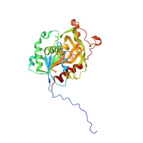
2VDJ - PubMed Abstract:
Bacteria and yeast rely on either homoserine transsuccinylase (HTS, metA) or homoserine transacetylase (HTA; met2) for the biosynthesis of methionine. Although HTS and HTA catalyze similar chemical reactions, these proteins are typically unrelated in both sequence and three-dimensional structure. Here we present the 2.0 A resolution x-ray crystal structure of the Bacillus cereus metA protein in complex with homoserine, which provides the first view of a ligand bound to either HTA or HTS. Surprisingly, functional analysis of the B. cereus metA protein shows that it does not use succinyl-CoA as a substrate. Instead, the protein catalyzes the transacetylation of homoserine using acetyl-CoA. Therefore, the B. cereus metA protein functions as an HTA despite greater than 50% sequence identity with bona fide HTS proteins. This result emphasizes the need for functional confirmation of annotations of enzyme function based on either sequence or structural comparisons. Kinetic analysis of site-directed mutants reveals that the B. cereus metA protein and the E. coli HTS share a common catalytic mechanism. Structural and functional examination of the B. cereus metA protein reveals that a single amino acid in the active site determines acetyl-CoA (Glu-111) versus succinyl-CoA (Gly-111) specificity in the metA-like of acyltransferases. Switching of this residue provides a mechanism for evolving substrate specificity in bacterial methionine biosynthesis. Within this enzyme family, HTS and HTA activity likely arises from divergent evolution in a common structural scaffold with conserved catalytic machinery and homoserine binding sites.
Organizational Affiliation:
Donald Danforth Plant Science Center, St. Louis, Missouri 63132, USA.