2VCY

Crystal Structure of 2-Enoyl Thioester Reductase of Human FAS II


Experimental Data Snapshot

  • Method: X-RAY DIFFRACTION
  • Resolution: 2.41 Å
  • R-Value Free: 0.267 
  • R-Value Work: 0.207 
  • R-Value Observed: 0.210 

wwPDB Validation   3D Report Full Report


This is version 1.4 of the entry. See complete history


Literature

Structural Enzymological Studies of 2-Enoyl Thioester Reductase of the Human Mitochondrial Fas II Pathway: New Insights Into its Substrate Recognition Properties.

Chen, Z.J.Pudas, R.Sharma, S.Smart, O.S.Juffer, A.H.Hiltunen, J.K.Wierenga, R.K.Haapalainen, A.M.

(2008) J Mol Biol 379: 830

  • DOI: https://doi.org/10.1016/j.jmb.2008.04.041
  • Primary Citation of Related Structures:  
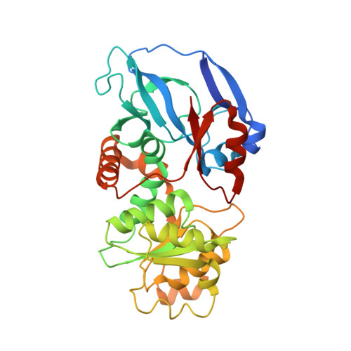
    2VCY

  • PubMed Abstract: 

    Structural and kinetic properties of the human 2-enoyl thioester reductase [mitochondrial enoyl-coenzyme A reductase (MECR)/ETR1] of the mitochondrial fatty acid synthesis (FAS) II pathway have been determined. The crystal structure of this dimeric enzyme (at 2.4 A resolution) suggests that the binding site for the recognition helix of the acyl carrier protein is in a groove between the two adjacent monomers. This groove is connected via the pantetheine binding cleft to the active site. The modeled mode of NADPH binding, using molecular dynamics calculations, suggests that Tyr94 and Trp311 are critical for catalysis, which is supported by enzyme kinetic data. A deep, water-filled pocket, shaped by hydrophobic and polar residues and extending away from the catalytic site, was recognized. This pocket can accommodate a fatty acyl tail of up to 16 carbons. Mutagenesis of the residues near the end of this pocket confirms the importance of this region for the binding of substrate molecules with long fatty acyl tails. Furthermore, the kinetic analysis of the wild-type MECR/ETR1 shows a bimodal distribution of catalytic efficiencies, in agreement with the notion that two major products are generated by the mitochondrial FAS II pathway.


  • Organizational Affiliation

    Biocenter Oulu and Department of Biochemistry, University of Oulu, P.O. Box 3000, FI-90014, Oulu, Finland.


Macromolecules
Find similar proteins by:  (by identity cutoff)  |  3D Structure
Entity ID: 1
MoleculeChains Sequence LengthOrganismDetailsImage
TRANS-2-ENOYL-COA REDUCTASE
A, B
344Homo sapiensMutation(s): 0 
EC: 1.3.1.38
UniProt & NIH Common Fund Data Resources
Find proteins for Q9BV79 (Homo sapiens)
Explore Q9BV79 
Go to UniProtKB:  Q9BV79
PHAROS:  Q9BV79
GTEx:  ENSG00000116353 
Entity Groups  
Sequence Clusters30% Identity50% Identity70% Identity90% Identity95% Identity100% Identity
UniProt GroupQ9BV79
Sequence Annotations
Expand
  • Reference Sequence
Small Molecules
Ligands 1 Unique
IDChains Name / Formula / InChI Key2D Diagram3D Interactions
SO4
Query on SO4

Download Ideal Coordinates CCD File 
C [auth A]
D [auth A]
E [auth A]
F [auth A]
G [auth A]
C [auth A],
D [auth A],
E [auth A],
F [auth A],
G [auth A],
H [auth B],
I [auth B],
J [auth B],
K [auth B],
L [auth B],
M [auth B]
SULFATE ION
O4 S
QAOWNCQODCNURD-UHFFFAOYSA-L
Experimental Data & Validation

Experimental Data

  • Method: X-RAY DIFFRACTION
  • Resolution: 2.41 Å
  • R-Value Free: 0.267 
  • R-Value Work: 0.207 
  • R-Value Observed: 0.210 
  • Space Group: P 42 21 2
Unit Cell:
Length ( Å )Angle ( ˚ )
a = 104.62α = 90
b = 104.62β = 90
c = 146.56γ = 90
Software Package:
Software NamePurpose
BUSTER-TNTrefinement
iMOSFLMdata reduction
SCALAdata scaling
PHASERphasing

Structure Validation

View Full Validation Report


Entry History 

Deposition Data

Revision History  (Full details and data files)

  • Version 1.0: 2008-06-03
    Type: Initial release
  • Version 1.1: 2011-05-08
    Changes: Version format compliance
  • Version 1.2: 2011-07-13
    Changes: Version format compliance
  • Version 1.3: 2019-07-24
    Changes: Data collection
  • Version 1.4: 2023-12-13
    Changes: Data collection, Database references, Other, Refinement description