2V9P

Crystal structure of papillomavirus E1 hexameric helicase DNA-free form


Experimental Data Snapshot

  • Method: X-RAY DIFFRACTION
  • Resolution: 3.00 Å
  • R-Value Free: 0.271 
  • R-Value Work: 0.219 
  • R-Value Observed: 0.219 

wwPDB Validation   3D Report Full Report


This is version 1.2 of the entry. See complete history


Literature

Papillomavirus E1 Helicase Assembly Maintains an Asymmetric State in the Absence of DNA and Nucleotide Cofactors.

Sanders, C.M.Kovalevskiy, O.V.Sizov, D.Lebedev, A.A.Isupov, M.N.Antson, A.A.

(2007) Nucleic Acids Res 35: 6451

  • DOI: https://doi.org/10.1093/nar/gkm705
  • Primary Citation of Related Structures:  
    2V9P

  • PubMed Abstract: 

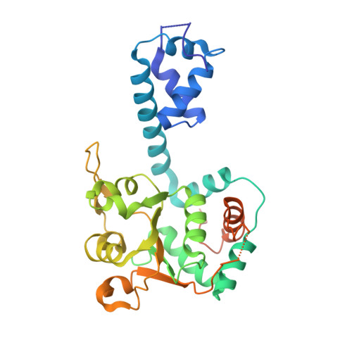
    Concerted, stochastic and sequential mechanisms of action have been proposed for different hexameric AAA+ molecular motors. Here we report the crystal structure of the E1 helicase from bovine papillomavirus, where asymmetric assembly is for the first time observed in the absence of nucleotide cofactors and DNA. Surprisingly, the ATP-binding sites adopt specific conformations linked to positional changes in the DNA-binding hairpins, which follow a wave-like trajectory, as observed previously in the E1/DNA/ADP complex. The protein's assembly thus maintains such an asymmetric state in the absence of DNA and nucleotide cofactors, allowing consideration of the E1 helicase action as the propagation of a conformational wave around the protein ring. The data imply that the wave's propagation within the AAA+ domains is not necessarily coupled with a strictly sequential hydrolysis of ATP. Since a single ATP hydrolysis event would affect the whole hexamer, such events may simply serve to rectify the direction of the wave's motion.


  • Organizational Affiliation

    Institute for Cancer Studies, University of Sheffield, Beech Hill Road, Sheffield, S10 2RX, UK.


Macromolecules
Find similar proteins by:  (by identity cutoff)  |  3D Structure
Entity ID: 1
MoleculeChains Sequence LengthOrganismDetailsImage
REPLICATION PROTEIN E1
A, B, C, D, E
A, B, C, D, E, F, G, H, I, J, K, L
305Deltapapillomavirus 4Mutation(s): 0 
EC: 3.6.1
UniProt
Find proteins for P03116 (Bovine papillomavirus type 1)
Explore P03116 
Go to UniProtKB:  P03116
Entity Groups  
Sequence Clusters30% Identity50% Identity70% Identity90% Identity95% Identity100% Identity
UniProt GroupP03116
Sequence Annotations
Expand
  • Reference Sequence
Small Molecules
Ligands 2 Unique
IDChains Name / Formula / InChI Key2D Diagram3D Interactions
PO4
Query on PO4

Download Ideal Coordinates CCD File 
BA [auth H]
CA [auth H]
EA [auth I]
FA [auth I]
HA [auth J]
BA [auth H],
CA [auth H],
EA [auth I],
FA [auth I],
HA [auth J],
IA [auth K],
JA [auth L],
M [auth A],
N [auth A],
P [auth B],
Q [auth B],
S [auth C],
T [auth C],
V [auth D],
W [auth E],
X [auth F],
Y [auth G],
Z [auth G]
PHOSPHATE ION
O4 P
NBIIXXVUZAFLBC-UHFFFAOYSA-K
MG
Query on MG

Download Ideal Coordinates CCD File 
AA [auth G]
DA [auth H]
GA [auth I]
O [auth A]
R [auth B]
AA [auth G],
DA [auth H],
GA [auth I],
O [auth A],
R [auth B],
U [auth C]
MAGNESIUM ION
Mg
JLVVSXFLKOJNIY-UHFFFAOYSA-N
Experimental Data & Validation

Experimental Data

  • Method: X-RAY DIFFRACTION
  • Resolution: 3.00 Å
  • R-Value Free: 0.271 
  • R-Value Work: 0.219 
  • R-Value Observed: 0.219 
  • Space Group: P 21 21 21
Unit Cell:
Length ( Å )Angle ( ˚ )
a = 135.111α = 90
b = 180.649β = 90
c = 187.533γ = 90
Software Package:
Software NamePurpose
REFMACrefinement
DENZOdata reduction
SCALEPACKdata scaling
MOLREPphasing

Structure Validation

View Full Validation Report


Entry History 

Deposition Data

Revision History  (Full details and data files)

  • Version 1.0: 2007-10-02
    Type: Initial release
  • Version 1.1: 2011-07-13
    Changes: Advisory, Refinement description, Version format compliance
  • Version 1.2: 2023-12-13
    Changes: Data collection, Database references, Derived calculations, Other, Refinement description