Molecular Cloning, Characterisation and Ligand- Bound Structure of an Azoreductase from Pseudomonas Aeruginosa
Wang, C.-J., Hagemeier, C., Rahman, N., Lowe, E.D., Noble, M.E.M., Coughtrie, M., Sim, E., Westwood, I.M.(2007) J Mol Biol 373: 1213
- PubMed: 17904577
- DOI: https://doi.org/10.1016/j.jmb.2007.08.048
- Primary Citation of Related Structures:
2V9C - PubMed Abstract:
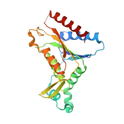
The gene PA0785 from Pseudomonas aeruginosa strain PAO1, which is annotated as a probable acyl carrier protein phosphodiesterase (acpD), has been cloned and heterologously overexpressed in Escherichia coli. The purified recombinant enzyme exhibits activity corresponding to that of azoreductase but not acpD. Each recombinant protein molecule has an estimated molecular mass of 23,050 Da and one non-covalently bound FMN as co-factor. This enzyme, now identified as azoreductase 1 from Pseudomonas aeruginosa (paAzoR1), is a flavodoxin-like protein with an apparent molecular mass of 110 kDa as determined by gel-filtration chromatography, indicating that the protein is likely to be tetrameric in solution. The three-dimensional structure of paAzoR1, in complex with the substrate methyl red, was solved at a resolution of 2.18 A by X-ray crystallography. The protein exists as a dimer of dimers in the crystal lattice, with two spatially separated active sites per dimer, and the active site of paAzoR1 was shown to be a well-conserved hydrophobic pocket formed between two monomers. The paAzoR1 enzyme is able to reduce different classes of azo dyes and activate several azo pro-drugs used in the treatment of inflammatory bowel disease (IBD). During azo reduction, FMN serves as a redox centre in the electron-transferring system by mediating the electron transfer from NAD(P)H to the azo substrate. The spectral properties of paAzoR1 demonstrate the hydrophobic interaction between FMN and the active site in the protein. The structure of the ligand-bound protein also highlights the pi-stacking interactions between FMN and the azo substrate.
Organizational Affiliation:
Department of Pharmacology, University of Oxford, Mansfield Road, Oxford, OX1 3QT, UK.