2V98

Structure of the complex of TcAChE with 1-(2-nitrophenyl)-2,2,2- trifluoroethyl-arsenocholine after a 9 seconds annealing to room temperature, during the first 5 seconds of which laser irradiation at 266nm took place


Experimental Data Snapshot

  • Method: X-RAY DIFFRACTION
  • Resolution: 3.00 Å
  • R-Value Free: 0.249 
  • R-Value Work: 0.202 
  • R-Value Observed: 0.202 

wwPDB Validation   3D Report Full Report


This is version 1.6 of the entry. See complete history


Literature

Use of a 'Caged' Analog to Study Traffic of Choline within Acetylcholinesterase by Kinetic Crystallography

Colletier, J.-P.Royant, A.Specht, A.Sanson, B.Nachon, F.Masson, P.Zaccai, G.Sussman, J.L.Goeldner, M.Silman, I.Bourgeois, D.Weik, M.

(2007) Acta Crystallogr D Biol Crystallogr 63: 1115

  • DOI: https://doi.org/10.1107/S0907444907044472
  • Primary Citation of Related Structures:  
    2V96, 2V97, 2V98, 2VA9

  • PubMed Abstract: 

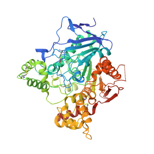
    Acetylcholinesterase plays a crucial role in nerve-impulse transmission at cholinergic synapses. The apparent paradox that it displays high turnover despite its active site being buried raises cogent questions as to how the traffic of substrates and products to and from the active site can occur so rapidly in such circumstances. Here, a kinetic crystallography strategy aimed at structurally addressing the issue of product traffic in acetylcholinesterase is presented, in which UV-laser-induced cleavage of a photolabile precursor of the enzymatic product analogue arsenocholine, 'caged' arsenocholine, is performed in a temperature-controlled X-ray crystallography regime. The 'caged' arsenocholine was shown to bind at both the active and peripheral sites of acetylcholinesterase. UV irradiation of a complex with acetylcholinesterase during a brief temperature excursion from 100 K to room temperature is most likely to have resulted in a decrease in occupancy by the caged compound. Microspectrophotometric experiments showed that the caged compound had indeed been photocleaved. It is proposed that a fraction of the arsenocholine molecules released within the crystal had been expelled from both the active and the peripheral sites. Partial q-weighted difference refinement revealed a relative movement of the two domains in acetylcholinesterase after photolysis and the room-temperature excursion, resulting in an increase in the active-site gorge volume of 30% and 35% in monomers A and B of the asymmetric unit, respectively. Moreover, an alternative route to the active-site gorge of the enzyme appeared to open. This structural characterization of acetylcholinesterase 'at work' is consistent with the idea that choline exits from the enzyme after catalysis either via the gorge or via an alternative 'backdoor' trajectory.


  • Organizational Affiliation

    Laboratoire de Biophysique Moléculaire, Institut de Biologie Structurale Jean-Pierre EBEL, UMR 5075 CNRS-CEA-UJF, 38027 Grenoble, France.


Macromolecules
Find similar proteins by:  (by identity cutoff)  |  3D Structure
Entity ID: 1
MoleculeChains Sequence LengthOrganismDetailsImage
ACETYLCHOLINESTERASE
A, B
537Tetronarce californicaMutation(s): 0 
EC: 3.1.1.7
UniProt
Find proteins for P04058 (Tetronarce californica)
Explore P04058 
Go to UniProtKB:  P04058
Entity Groups  
Sequence Clusters30% Identity50% Identity70% Identity90% Identity95% Identity100% Identity
UniProt GroupP04058
Sequence Annotations
Expand
  • Reference Sequence
Small Molecules
Ligands 3 Unique
IDChains Name / Formula / InChI Key2D Diagram3D Interactions
CFQ
Query on CFQ

Download Ideal Coordinates CCD File 
D [auth A],
E [auth A],
H [auth B],
I [auth B]
1-(2-nitrophenyl)-2,2,2-trifluoroethyl]-arsenocholine
C13 H19 As F3 N O3
LUSVMAVUPPIHKA-LBPRGKRZSA-N
NAG
Query on NAG

Download Ideal Coordinates CCD File 
F [auth A],
G [auth A],
J [auth B],
K [auth B]
2-acetamido-2-deoxy-beta-D-glucopyranose
C8 H15 N O6
OVRNDRQMDRJTHS-FMDGEEDCSA-N
CL
Query on CL

Download Ideal Coordinates CCD File 
C [auth A]CHLORIDE ION
Cl
VEXZGXHMUGYJMC-UHFFFAOYSA-M
Experimental Data & Validation

Experimental Data

  • Method: X-RAY DIFFRACTION
  • Resolution: 3.00 Å
  • R-Value Free: 0.249 
  • R-Value Work: 0.202 
  • R-Value Observed: 0.202 
  • Space Group: P 21 21 21
Unit Cell:
Length ( Å )Angle ( ˚ )
a = 91.62α = 90
b = 104.47β = 90
c = 148.8γ = 90
Software Package:
Software NamePurpose
CNSrefinement
XDSdata reduction
XSCALEdata scaling
CNSphasing

Structure Validation

View Full Validation Report


Entry History 

Deposition Data

Revision History  (Full details and data files)

  • Version 1.0: 2007-11-20
    Type: Initial release
  • Version 1.1: 2011-05-08
    Changes: Version format compliance
  • Version 1.2: 2011-07-13
    Changes: Version format compliance
  • Version 1.3: 2019-04-03
    Changes: Data collection, Experimental preparation, Other
  • Version 1.4: 2020-07-29
    Type: Remediation
    Reason: Carbohydrate remediation
    Changes: Data collection, Derived calculations, Other, Structure summary
  • Version 1.5: 2021-05-12
    Changes: Derived calculations, Structure summary
  • Version 1.6: 2023-12-13
    Changes: Data collection, Database references, Refinement description