2V6Y

Structure of the MIT domain from a S. solfataricus Vps4-like ATPase


Experimental Data Snapshot

  • Method: X-RAY DIFFRACTION
  • Resolution: 2.40 Å
  • R-Value Free: 0.277 
  • R-Value Work: 0.252 
  • R-Value Observed: 0.253 

wwPDB Validation   3D Report Full Report


Ligand Structure Quality Assessment 


This is version 1.1 of the entry. See complete history


Literature

Structural Basis for Selective Recognition of Escrt-III by the Aaa ATPase Vps4

Obita, T.Saksena, S.Ghazi-Tabatabai, S.Gill, D.J.Perisic, O.Emr, S.D.Williams, R.L.

(2007) Nature 449: 735

  • DOI: https://doi.org/10.1038/nature06171
  • Primary Citation of Related Structures:  
    2V6X, 2V6Y

  • PubMed Abstract: 

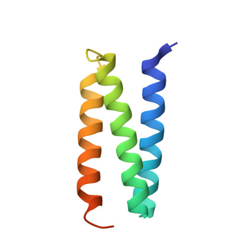
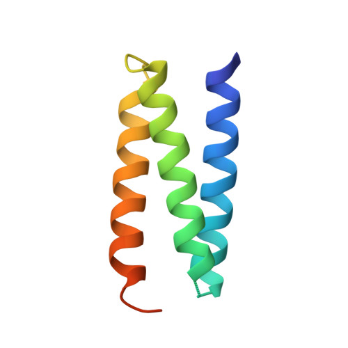
    The AAA+ ATPases are essential for various activities such as membrane trafficking, organelle biogenesis, DNA replication, intracellular locomotion, cytoskeletal remodelling, protein folding and proteolysis. The AAA ATPase Vps4, which is central to endosomal traffic to lysosomes, retroviral budding and cytokinesis, dissociates ESCRT complexes (the endosomal sorting complexes required for transport) from membranes. Here we show that, of the six ESCRT--related subunits in yeast, only Vps2 and Did2 bind the MIT (microtubule interacting and transport) domain of Vps4, and that the carboxy-terminal 30 residues of the subunits are both necessary and sufficient for interaction. We determined the crystal structure of the Vps2 C terminus in a complex with the Vps4 MIT domain, explaining the basis for selective ESCRT-III recognition. MIT helices alpha2 and alpha3 recognize a (D/E)xxLxxRLxxL(K/R) motif, and mutations within this motif cause sorting defects in yeast. Our crystal structure of the amino-terminal domain of an archaeal AAA ATPase of unknown function shows that it is closely related to the MIT domain of Vps4. The archaeal ATPase interacts with an archaeal ESCRT-III-like protein even though these organisms have no endomembrane system, suggesting that the Vps4/ESCRT-III partnership is a relic of a function that pre-dates the divergence of eukaryotes and Archaea.


  • Organizational Affiliation

    MRC Laboratory of Molecular Biology, Medical Research Council Centre, Cambridge CB2 0QH, UK.


Macromolecules
Find similar proteins by:  (by identity cutoff)  |  3D Structure
Entity ID: 1
MoleculeChains Sequence LengthOrganismDetailsImage
AAA FAMILY ATPASE, P60 KATANIN83Saccharolobus solfataricusMutation(s): 0 
EC: 3.6.4.6
UniProt
Find proteins for Q97ZJ7 (Saccharolobus solfataricus (strain ATCC 35092 / DSM 1617 / JCM 11322 / P2))
Explore Q97ZJ7 
Go to UniProtKB:  Q97ZJ7
Entity Groups  
Sequence Clusters30% Identity50% Identity70% Identity90% Identity95% Identity100% Identity
UniProt GroupQ97ZJ7
Sequence Annotations
Expand
  • Reference Sequence
Find similar proteins by:  (by identity cutoff)  |  3D Structure
Entity ID: 2
MoleculeChains Sequence LengthOrganismDetailsImage
AAA FAMILY ATPASE, P60 KATANIN83Saccharolobus solfataricusMutation(s): 0 
EC: 3.6.4.6
UniProt
Find proteins for Q97ZJ7 (Saccharolobus solfataricus (strain ATCC 35092 / DSM 1617 / JCM 11322 / P2))
Explore Q97ZJ7 
Go to UniProtKB:  Q97ZJ7
Entity Groups  
Sequence Clusters30% Identity50% Identity70% Identity90% Identity95% Identity100% Identity
UniProt GroupQ97ZJ7
Sequence Annotations
Expand
  • Reference Sequence
Small Molecules
Experimental Data & Validation

Experimental Data

  • Method: X-RAY DIFFRACTION
  • Resolution: 2.40 Å
  • R-Value Free: 0.277 
  • R-Value Work: 0.252 
  • R-Value Observed: 0.253 
  • Space Group: I 21 21 21
Unit Cell:
Length ( Å )Angle ( ˚ )
a = 56.178α = 90
b = 69.267β = 90
c = 123.184γ = 90
Software Package:
Software NamePurpose
MOSFLMdata reduction
SCALAdata scaling
SnBphasing
autoSHARPphasing
REFMACrefinement

Structure Validation

View Full Validation Report


Ligand Structure Quality Assessment 


Entry History 

Deposition Data

Revision History  (Full details and data files)

  • Version 1.0: 2007-10-16
    Type: Initial release
  • Version 1.1: 2011-07-13
    Changes: Advisory, Refinement description, Version format compliance