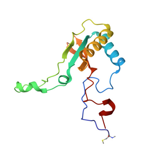
2V68

Crystal structure of Chlamydomonas reinhardtii Rubisco with large- subunit mutations V331A, T342I


Experimental Data Snapshot

  • Method: X-RAY DIFFRACTION
  • Resolution: 2.30 Å
  • R-Value Free: 0.203 
  • R-Value Work: 0.172 
  • R-Value Observed: 0.174 

wwPDB Validation   3D Report Full Report


Ligand Structure Quality Assessment 


This is version 1.4 of the entry. See complete history


Literature

Structural Analysis of Altered Large-Subunit Loop-6-Carboxy-Terminus Interactions that Influence Catalytic Efficiency and Co2 O2 Specificity of Ribulose-1,5-Bisphosphate Carboxylase Oxygenase

Karkehabadi, S.Satagopan, S.Taylor, T.C.Spreitzer, R.J.Andersson, I.

(2007) Biochemistry 46: 11080

  • DOI: https://doi.org/10.1021/bi701063f
  • Primary Citation of Related Structures:  
    2V63, 2V67, 2V68, 2V69, 2V6A

  • PubMed Abstract: 

    The loop between alpha-helix 6 and beta-strand 6 in the alpha/beta-barrel of ribulose-1,5-bisphosphate carboxylase/oxygenase plays a key role in discriminating between CO2 and O2. Genetic screening in Chlamydomonas reinhardtii previously identified a loop-6 V331A substitution that decreases carboxylation and CO2/O2 specificity. Revertant selection identified T342I and G344S substitutions that restore photosynthetic growth by increasing carboxylation and specificity of the V331A enzyme. In numerous X-ray crystal structures, loop 6 is closed or open depending on the activation state of the enzyme and the presence or absence of ligands. The carboxy terminus folds over loop 6 in the closed state. To study the molecular basis for catalysis, directed mutagenesis and chloroplast transformation were used to create T342I and G344S substitutions alone. X-ray crystal structures were then solved for the V331A, V331A/T342I, T342I, and V331A/G344S enzymes, as well as for a D473E enzyme created to assess the role of the carboxy terminus in loop-6 closure. V331A disturbs a hydrophobic pocket, abolishing several van der Waals interactions. These changes are complemented by T342I and G344S, both of which alone cause decreases in CO2/O2 specificity. In the V331A/T342I revertant enzyme, Arg339 main-chain atoms are displaced. In V331A/G344S, alpha-helix 6 is shifted. D473E causes disorder of the carboxy terminus, but loop 6 remains closed. Interactions between a transition-state analogue and several residues are altered in the mutant enzymes. However, active-site Lys334 at the apex of loop 6 has a normal conformation. A variety of subtle interactions must be responsible for catalytic efficiency and CO2/O2 specificity.


  • Organizational Affiliation

    Department of Molecular Biology, Swedish University of Agricultural Sciences, BMC Box 590, 751 24 Uppsala, Sweden.


Macromolecules
Find similar proteins by:  (by identity cutoff)  |  3D Structure
Entity ID: 1
MoleculeChains Sequence LengthOrganismDetailsImage
RIBULOSE BISPHOSPHATE CARBOXYLASE LARGE CHAIN
A, B, C, D, E
A, B, C, D, E, F, G, H
475Chlamydomonas reinhardtiiMutation(s): 2 
EC: 4.1.1.39
UniProt
Find proteins for P00877 (Chlamydomonas reinhardtii)
Explore P00877 
Go to UniProtKB:  P00877
Entity Groups  
Sequence Clusters30% Identity50% Identity70% Identity90% Identity95% Identity100% Identity
UniProt GroupP00877
Sequence Annotations
Expand
  • Reference Sequence
Find similar proteins by:  (by identity cutoff)  |  3D Structure
Entity ID: 2
MoleculeChains Sequence LengthOrganismDetailsImage
RIBULOSE BISPHOSPHATE CARBOXYLASE SMALL CHAIN 1
I, J, K, L, M
I, J, K, L, M, N, O, P
140Chlamydomonas reinhardtiiMutation(s): 0 
EC: 4.1.1.39
UniProt
Find proteins for P00873 (Chlamydomonas reinhardtii)
Explore P00873 
Go to UniProtKB:  P00873
Entity Groups  
Sequence Clusters30% Identity50% Identity70% Identity90% Identity95% Identity100% Identity
UniProt GroupP00873
Sequence Annotations
Expand
  • Reference Sequence
Small Molecules
Ligands 3 Unique
IDChains Name / Formula / InChI Key2D Diagram3D Interactions
CAP
Query on CAP

Download Ideal Coordinates CCD File 
EA [auth C]
EB [auth G]
JB [auth H]
LA [auth D]
R [auth A]
EA [auth C],
EB [auth G],
JB [auth H],
LA [auth D],
R [auth A],
RA [auth E],
XA [auth F],
Y [auth B]
2-CARBOXYARABINITOL-1,5-DIPHOSPHATE
C6 H14 O13 P2
ITHCSGCUQDMYAI-ZMIZWQJLSA-N
EDO
Query on EDO

Download Ideal Coordinates CCD File 
AA [auth B]
AB [auth F]
AC [auth O]
BA [auth B]
BB [auth F]
AA [auth B],
AB [auth F],
AC [auth O],
BA [auth B],
BB [auth F],
BC [auth O],
CA [auth B],
CB [auth F],
CC [auth P],
FA [auth C],
FB [auth G],
GA [auth C],
GB [auth G],
HA [auth C],
HB [auth G],
IA [auth C],
JA [auth C],
KB [auth H],
LB [auth H],
MA [auth D],
MB [auth H],
NA [auth D],
NB [auth H],
OA [auth D],
OB [auth H],
PA [auth D],
PB [auth H],
QB [auth I],
RB [auth I],
S [auth A],
SA [auth E],
SB [auth J],
T [auth A],
TA [auth E],
TB [auth J],
U [auth A],
UA [auth E],
UB [auth K],
V [auth A],
VA [auth F],
VB [auth K],
W [auth A],
WB [auth L],
XB [auth M],
YA [auth F],
YB [auth N],
Z [auth B],
ZA [auth F],
ZB [auth N]
1,2-ETHANEDIOL
C2 H6 O2
LYCAIKOWRPUZTN-UHFFFAOYSA-N
MG
Query on MG

Download Ideal Coordinates CCD File 
DA [auth C]
DB [auth G]
IB [auth H]
KA [auth D]
Q [auth A]
DA [auth C],
DB [auth G],
IB [auth H],
KA [auth D],
Q [auth A],
QA [auth E],
WA [auth F],
X [auth B]
MAGNESIUM ION
Mg
JLVVSXFLKOJNIY-UHFFFAOYSA-N
Modified Residues  4 Unique
IDChains TypeFormula2D DiagramParent
HYP
Query on HYP
A, B, C, D, E
A, B, C, D, E, F, G, H
L-PEPTIDE LINKINGC5 H9 N O3PRO
KCX
Query on KCX
A, B, C, D, E
A, B, C, D, E, F, G, H
L-PEPTIDE LINKINGC7 H14 N2 O4LYS
SMC
Query on SMC
A, B, C, D, E
A, B, C, D, E, F, G, H
L-PEPTIDE LINKINGC4 H9 N O2 SCYS
MME
Query on MME
I, J, K, L, M
I, J, K, L, M, N, O, P
L-PEPTIDE LINKINGC6 H13 N O2 SMET
Experimental Data & Validation

Experimental Data

  • Method: X-RAY DIFFRACTION
  • Resolution: 2.30 Å
  • R-Value Free: 0.203 
  • R-Value Work: 0.172 
  • R-Value Observed: 0.174 
  • Space Group: P 1 21 1
Unit Cell:
Length ( Å )Angle ( ˚ )
a = 120.421α = 90
b = 178.236β = 117.72
c = 122.758γ = 90
Software Package:
Software NamePurpose
REFMACrefinement
DENZOdata reduction
SCALEPACKdata scaling
MOLREPphasing

Structure Validation

View Full Validation Report


Ligand Structure Quality Assessment 


Entry History 

Deposition Data

Revision History  (Full details and data files)

  • Version 1.0: 2007-08-07
    Type: Initial release
  • Version 1.1: 2011-05-08
    Changes: Version format compliance
  • Version 1.2: 2011-07-13
    Changes: Version format compliance
  • Version 1.3: 2018-01-17
    Changes: Data collection
  • Version 1.4: 2023-12-13
    Changes: Data collection, Database references, Derived calculations, Other, Refinement description