2V57

Crystal structure of the TetR-like transcriptional regulator LfrR from Mycobacterium smegmatis in complex with proflavine


Experimental Data Snapshot

  • Method: X-RAY DIFFRACTION
  • Resolution: 1.90 Å
  • R-Value Free: 0.234 
  • R-Value Work: 0.196 
  • R-Value Observed: 0.198 

wwPDB Validation   3D Report Full Report


Ligand Structure Quality Assessment 


This is version 1.2 of the entry. See complete history


Literature

Structural Plasticity and Distinct Drug-Binding Modes of Lfrr, a Mycobacterial Efflux Pump Regulator.

Bellinzoni, M.Buroni, S.Schaeffer, F.Riccardi, G.De Rossi, E.Alzari, P.M.

(2009) J Bacteriol 191: 7531

  • DOI: https://doi.org/10.1128/JB.00631-09
  • Primary Citation of Related Structures:  
    2V57, 2WGB

  • PubMed Abstract: 

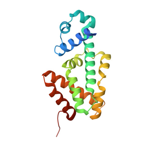
    The TetR-like transcriptional repressor LfrR controls the expression of the gene encoding the Mycobacterium smegmatis efflux pump LfrA, which actively extrudes fluoroquinolones, cationic dyes, and anthracyclines from the cell and promotes intrinsic antibiotic resistance. The crystal structure of the apoprotein form of the repressor reveals a structurally asymmetric homodimer exhibiting local unfolding and a blocked drug-binding site, emphasizing the significant conformational plasticity of the protein necessary for DNA and multidrug recognition. Crystallographic and calorimetric studies of LfrR-drug complexes further confirm the intrinsic flexibility of the homodimer, which provides a dynamic mechanism to broaden multidrug binding specificity and may be a general property of transcriptional repressors regulating microbial efflux pump expression.


  • Organizational Affiliation

    Institut Pasteur, Unité de Biochimie Structurale and CNRS URA 2185, Institut Pasteur, 75724 Paris Cedex 15, France.


Macromolecules
Find similar proteins by:  (by identity cutoff)  |  3D Structure
Entity ID: 1
MoleculeChains Sequence LengthOrganismDetailsImage
TETR FAMILY TRANSCRIPTIONAL REPRESSOR LFRR
A, B, C, D
190Mycolicibacterium smegmatisMutation(s): 0 
UniProt
Find proteins for Q58L87 (Mycolicibacterium smegmatis)
Explore Q58L87 
Go to UniProtKB:  Q58L87
Entity Groups  
Sequence Clusters30% Identity50% Identity70% Identity90% Identity95% Identity100% Identity
UniProt GroupQ58L87
Sequence Annotations
Expand
  • Reference Sequence
Small Molecules
Ligands 3 Unique
IDChains Name / Formula / InChI Key2D Diagram3D Interactions
PRL
Query on PRL

Download Ideal Coordinates CCD File 
E [auth A],
I [auth C]
PROFLAVIN
C13 H11 N3
WDVSHHCDHLJJJR-UHFFFAOYSA-N
SO4
Query on SO4

Download Ideal Coordinates CCD File 
G [auth B]
H [auth B]
J [auth C]
L [auth D]
M [auth D]
G [auth B],
H [auth B],
J [auth C],
L [auth D],
M [auth D],
N [auth D]
SULFATE ION
O4 S
QAOWNCQODCNURD-UHFFFAOYSA-L
IPA
Query on IPA

Download Ideal Coordinates CCD File 
F [auth A],
K [auth C]
ISOPROPYL ALCOHOL
C3 H8 O
KFZMGEQAYNKOFK-UHFFFAOYSA-N
Binding Affinity Annotations 
IDSourceBinding Affinity
PRL Binding MOAD:  2V57 Kd: 79 (nM) from 1 assay(s)
PDBBind:  2V57 Kd: 79 (nM) from 1 assay(s)
Experimental Data & Validation

Experimental Data

  • Method: X-RAY DIFFRACTION
  • Resolution: 1.90 Å
  • R-Value Free: 0.234 
  • R-Value Work: 0.196 
  • R-Value Observed: 0.198 
  • Space Group: P 1
Unit Cell:
Length ( Å )Angle ( ˚ )
a = 48.3α = 63.88
b = 62.44β = 88.34
c = 70.64γ = 88.13
Software Package:
Software NamePurpose
PHENIXrefinement
XDSdata reduction
SCALAdata scaling
MOLREPphasing

Structure Validation

View Full Validation Report


Ligand Structure Quality Assessment 


Entry History 

Deposition Data

Revision History  (Full details and data files)

  • Version 1.0: 2009-10-20
    Type: Initial release
  • Version 1.1: 2011-05-08
    Changes: Version format compliance
  • Version 1.2: 2011-07-13
    Changes: Version format compliance