Combining Crystallography and Molecular Dynamics: The Case of Schistosoma Mansoni Phospholipid Glutathione Peroxidase.
Dimastrogiovanni, D., Anselmi, M., Miele, A.E., Boumis, G., Petersson, L., Angelucci, F., Nola, A.D., Brunori, M., Bellelli, A.(2010) Proteins 78: 259
- PubMed: 19714775
- DOI: https://doi.org/10.1002/prot.22536
- Primary Citation of Related Structures:
2V1M, 2WGR - PubMed Abstract:
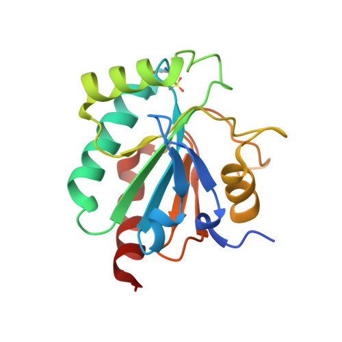
Oxidative stress is a widespread challenge for living organisms, and especially so for parasitic ones, given the fact that their hosts can produce reactive oxygen species (ROS) as a mechanism of defense. Thus, long lived parasites, such as the flatworm Schistosomes, have evolved refined enzymatic systems capable of detoxifying ROS. Among these, glutathione peroxidases (Gpx) are a family of sulfur or selenium-dependent isozymes sharing the ability to reduce peroxides using the reducing equivalents provided by glutathione or possibly small proteins such as thioredoxin. As for other frontline antioxidant enzymatic systems, Gpxs are localized in the tegument of the Schistosomes, the outermost defense layer. In this article, we present the first crystal structure at 1.0 and 1.7 A resolution of two recombinant SmGpxs, carrying the active site mutations Sec43Cys and Sec43Ser, respectively. The structures confirm that this enzyme belongs to the monomeric class 4 (phospholipid hydroperoxide) Gpx. In the case of the Sec to Cys mutant, the catalytic Cys residue is oxidized to sulfonic acid. By combining static crystallography with molecular dynamics simulations, we obtained insight into the substrate binding sites and the conformational changes relevant to catalysis, proposing a role for the unusual reactivity of the catalytic residue.
Organizational Affiliation:
Dipartimento di Scienze Biochimiche A. Rossi Fanelli and Istituto Pasteur, Fondazione Cenci Bolognetti, Sapienza University of Rome, Rome, Italy.