2UYT

Structure of L-rhamnulose kinase in complex with ADP and beta-L- rhamnulose.


Experimental Data Snapshot

  • Method: X-RAY DIFFRACTION
  • Resolution: 1.55 Å
  • R-Value Free: 0.209 
  • R-Value Work: 0.179 
  • R-Value Observed: 0.181 

wwPDB Validation   3D Report Full Report


Ligand Structure Quality Assessment 


This is version 1.4 of the entry. See complete history


Literature

Substrate Spectrum of L-Rhamnulose Kinase Related to Models Derived from Two Ternary Complex Structures.

Grueninger, D.Schulz, G.E.

(2007) FEBS Lett 581: 3127

  • DOI: https://doi.org/10.1016/j.febslet.2007.05.075
  • Primary Citation of Related Structures:  
    2UYT

  • PubMed Abstract: 

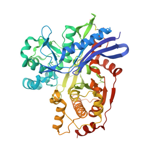
    The enzyme L-rhamnulose kinase from Escherichia coli participates in the degradation pathway of L-rhamnose, a common natural deoxy-hexose. The structure of the enzyme in a ternary complex with its substrates ADP and L-rhamnulose has been determined at 1.55A resolution and refined to R(cryst)/R(free) values of 0.179/0.209. The result was compared with the lower resolution structure of a corresponding complex containing L-fructose instead of L-rhamnulose. In light of the two established sugar positions and conformations, a number of rare sugars have been modeled into the active center of L-rhamnulose kinase and the model structures have been compared with the known enzymatic phosphorylation rates. Rare sugars are of rising interest for the synthesis of bioactive compounds.


  • Organizational Affiliation

    Institut für Organische Chemie und Biochemie, Albert-Ludwigs-Universität, Albertstrasse 21, 79104 Freiburg im Breisgau, Germany.


Macromolecules
Find similar proteins by:  (by identity cutoff)  |  3D Structure
Entity ID: 1
MoleculeChains Sequence LengthOrganismDetailsImage
RHAMNULOKINASE489Escherichia coli BL21(DE3)Mutation(s): 3 
EC: 2.7.1.5
UniProt
Find proteins for Q1R415 (Escherichia coli (strain UTI89 / UPEC))
Explore Q1R415 
Go to UniProtKB:  Q1R415
Entity Groups  
Sequence Clusters30% Identity50% Identity70% Identity90% Identity95% Identity100% Identity
UniProt GroupQ1R415
Sequence Annotations
Expand
  • Reference Sequence
Small Molecules
Ligands 2 Unique
IDChains Name / Formula / InChI Key2D Diagram3D Interactions
ADP
Query on ADP

Download Ideal Coordinates CCD File 
C [auth A]ADENOSINE-5'-DIPHOSPHATE
C10 H15 N5 O10 P2
XTWYTFMLZFPYCI-KQYNXXCUSA-N
LRH
Query on LRH

Download Ideal Coordinates CCD File 
B [auth A]6-deoxy-beta-L-fructofuranose
C6 H12 O5
CJJCPDZKQKUXSS-AZGQCCRYSA-N
Experimental Data & Validation

Experimental Data

  • Method: X-RAY DIFFRACTION
  • Resolution: 1.55 Å
  • R-Value Free: 0.209 
  • R-Value Work: 0.179 
  • R-Value Observed: 0.181 
  • Space Group: P 21 21 21
Unit Cell:
Length ( Å )Angle ( ˚ )
a = 51.043α = 90
b = 51.093β = 90
c = 158.85γ = 90
Software Package:
Software NamePurpose
REFMACrefinement
XDSdata reduction
XSCALEdata scaling
PHASERphasing

Structure Validation

View Full Validation Report


Ligand Structure Quality Assessment 


Entry History 

Deposition Data

Revision History  (Full details and data files)

  • Version 1.0: 2007-06-26
    Type: Initial release
  • Version 1.1: 2011-07-13
    Changes: Advisory, Version format compliance
  • Version 1.2: 2018-10-24
    Changes: Data collection, Source and taxonomy
  • Version 1.3: 2020-07-29
    Type: Remediation
    Reason: Carbohydrate remediation
    Changes: Data collection, Derived calculations, Other, Structure summary
  • Version 1.4: 2023-12-13
    Changes: Data collection, Database references, Refinement description, Structure summary