2UXQ

Isocitrate dehydrogenase from the psychrophilic bacterium Desulfotalea psychrophila: biochemical properties and crystal structure analysis


Experimental Data Snapshot

  • Method: X-RAY DIFFRACTION
  • Resolution: 1.75 Å
  • R-Value Free: 0.189 
  • R-Value Work: 0.146 
  • R-Value Observed: 0.146 

wwPDB Validation   3D Report Full Report


This is version 1.3 of the entry. See complete history


Literature

Structural and Functional Properties of Isocitrate Dehydrogenase from the Psychrophilic Bacterium Desulfotalea Psychrophila Reveal a Cold -Active Enzyme with an Unusual High Thermal

Fedoy, A.-E.Yang, N.Martinez, A.Leiros, H.-K.S.Steen, I.H.

(2007) J Mol Biol 372: 130

  • DOI: https://doi.org/10.1016/j.jmb.2007.06.040
  • Primary Citation of Related Structures:  
    2UXQ, 2UXR

  • PubMed Abstract: 

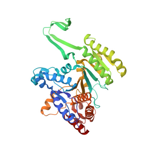
    Isocitrate dehydrogenase (IDH) has been studied extensively due to its central role in the Krebs cycle, catalyzing the oxidative NAD(P)(+)-dependent decarboxylation of isocitrate to alpha-ketoglutarate and CO(2). Here, we present the first crystal structure of IDH from a psychrophilic bacterium, Desulfotalea psychrophila (DpIDH). The structural information is combined with a detailed biochemical characterization and a comparative study with IDHs from the mesophilic bacterium Desulfitobacterium hafniense (DhIDH), porcine (PcIDH), human cytosolic (HcIDH) and the hyperthermophilic Thermotoga maritima (TmIDH). DpIDH was found to have a higher melting temperature (T(m)=66.9 degrees C) than its mesophilic homologues and a suboptimal catalytic efficiency at low temperatures. The thermodynamic activation parameters indicated a disordered active site, as seen also for the drastic increase in K(m) for isocitrate at elevated temperatures. A methionine cluster situated at the dimeric interface between the two active sites and a cluster of destabilizing charged amino acids in a region close to the active site might explain the poor isocitrate affinity. On the other hand, DpIDH was optimized for interacting with NADP(+) and the crystal structure revealed unique interactions with the cofactor. The highly acidic surface, destabilizing charged residues, fewer ion pairs and reduced size of ionic networks in DpIDH suggest a flexible global structure. However, strategic placement of ionic interactions stabilizing the N and C termini, and additional ionic interactions in the clasp domain as well as two enlarged aromatic clusters might counteract the destabilizing interactions and promote the increased thermal stability. The structure analysis of DpIDH illustrates how psychrophilic enzymes can adjust their flexibility in dynamic regions during their catalytic cycle without compromising the global stability of the protein.


  • Organizational Affiliation

    Department of Biology, University of Bergen, P.O. Box 7800, Jahnebakken 5, N-5020 Bergen, Norway.


Macromolecules
Find similar proteins by:  (by identity cutoff)  |  3D Structure
Entity ID: 1
MoleculeChains Sequence LengthOrganismDetailsImage
ISOCITRATE DEHYDROGENASE NATIVE
A, B, C, D
402Desulfotalea psychrophilaMutation(s): 0 
UniProt
Find proteins for Q6AQ66 (Desulfotalea psychrophila (strain LSv54 / DSM 12343))
Explore Q6AQ66 
Go to UniProtKB:  Q6AQ66
Entity Groups  
Sequence Clusters30% Identity50% Identity70% Identity90% Identity95% Identity100% Identity
UniProt GroupQ6AQ66
Sequence Annotations
Expand
  • Reference Sequence
Small Molecules
Ligands 4 Unique
IDChains Name / Formula / InChI Key2D Diagram3D Interactions
PEG
Query on PEG

Download Ideal Coordinates CCD File 
J [auth A],
Q [auth B],
R [auth B],
X [auth C],
Y [auth C]
DI(HYDROXYETHYL)ETHER
C4 H10 O3
MTHSVFCYNBDYFN-UHFFFAOYSA-N
SO4
Query on SO4

Download Ideal Coordinates CCD File 
CA [auth D]
DA [auth D]
H [auth A]
L [auth B]
M [auth B]
CA [auth D],
DA [auth D],
H [auth A],
L [auth B],
M [auth B],
N [auth B],
O [auth B],
P [auth B],
V [auth C],
W [auth C]
SULFATE ION
O4 S
QAOWNCQODCNURD-UHFFFAOYSA-L
GOL
Query on GOL

Download Ideal Coordinates CCD File 
AA [auth D]
BA [auth D]
E [auth A]
F [auth A]
G [auth A]
AA [auth D],
BA [auth D],
E [auth A],
F [auth A],
G [auth A],
K [auth B],
S [auth C],
T [auth C],
U [auth C],
Z [auth D]
GLYCEROL
C3 H8 O3
PEDCQBHIVMGVHV-UHFFFAOYSA-N
MG
Query on MG

Download Ideal Coordinates CCD File 
I [auth A]MAGNESIUM ION
Mg
JLVVSXFLKOJNIY-UHFFFAOYSA-N
Experimental Data & Validation

Experimental Data

  • Method: X-RAY DIFFRACTION
  • Resolution: 1.75 Å
  • R-Value Free: 0.189 
  • R-Value Work: 0.146 
  • R-Value Observed: 0.146 
  • Space Group: P 1
Unit Cell:
Length ( Å )Angle ( ˚ )
a = 59.324α = 98.9
b = 73.279β = 98.98
c = 126.413γ = 113.88
Software Package:
Software NamePurpose
REFMACrefinement
MOSFLMdata reduction
SCALAdata scaling
MOLREPphasing

Structure Validation

View Full Validation Report


Entry History 

Deposition Data

Revision History  (Full details and data files)

  • Version 1.0: 2008-02-05
    Type: Initial release
  • Version 1.1: 2011-05-08
    Changes: Version format compliance
  • Version 1.2: 2011-07-13
    Changes: Version format compliance
  • Version 1.3: 2023-12-13
    Changes: Data collection, Database references, Derived calculations, Other, Refinement description