2UX8

Crystal Structure of Sphingomonas elodea ATCC 31461 Glucose-1- phosphate uridylyltransferase in Complex with glucose-1-phosphate.


Experimental Data Snapshot

  • Method: X-RAY DIFFRACTION
  • Resolution: 2.65 Å
  • R-Value Free: 0.295 
  • R-Value Work: 0.245 
  • R-Value Observed: 0.245 

wwPDB Validation   3D Report Full Report


Ligand Structure Quality Assessment 


This is version 1.3 of the entry. See complete history


Literature

The Complex of Sphingomonas Elodea Atcc 31461 Glucose-1-Phosphate Uridylyltransferase with Glucose-1-Phosphate Reveals a Novel Quaternary Structure, Unique Among Nucleoside Diphosphate-Sugar Pyrophosphorylase Members.

Aragao, D.Fialho, A.M.Marques, A.R.Mitchell, E.P.Sa-Correia, I.Frazao, C.

(2007) J Bacteriol 189: 4520

  • DOI: https://doi.org/10.1128/JB.00277-07
  • Primary Citation of Related Structures:  
    2UX8

  • PubMed Abstract: 

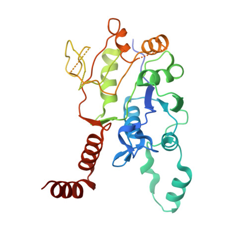
    Gellan gum is a widely used commercial material, available in many different forms. Its economic importance has led to studies into the biosynthesis of exopolysaccharide gellan gum, which is industrially prepared in high yields using Sphingomonas elodea ATCC 31461. Glucose-1-phosphate uridylyltransferase mediates the reversible conversion of glucose-1-phosphate and UTP into UDP-glucose and pyrophosphate, which is a key step in the biosynthetic pathway of gellan gums. Here we present the X-ray crystal structure of the glucose-1-phosphate uridylyltransferase from S. elodea. The S. elodea enzyme shares strong monomeric similarity with glucose-1-phosphate thymidylyltransferase, several structures of which are known, although the quaternary structures of the active enzymes are rather different. A detailed comparison between S. elodea glucose-1-phosphate uridylyltransferase and available thymidylyltransferases is described and shows remarkable structural similarities, despite the low sequence identities between the two divergent groups of proteins.


  • Organizational Affiliation

    Instituto de Tecnologia Química e Biológica, Universidade Nova de Lisboa, Apartado 127, 2781-901 Oeiras, Portugal.


Macromolecules
Find similar proteins by:  (by identity cutoff)  |  3D Structure
Entity ID: 1
MoleculeChains Sequence LengthOrganismDetailsImage
GLUCOSE-1-PHOSPHATE URIDYLYLTRANSFERASE
A, B, C, D, E
A, B, C, D, E, F, G, H
297Sphingomonas elodeaMutation(s): 0 
EC: 2.7.7.9
UniProt
Find proteins for Q5FYV5 (Sphingomonas elodea)
Explore Q5FYV5 
Go to UniProtKB:  Q5FYV5
Entity Groups  
Sequence Clusters30% Identity50% Identity70% Identity90% Identity95% Identity100% Identity
UniProt GroupQ5FYV5
Sequence Annotations
Expand
  • Reference Sequence
Experimental Data & Validation

Experimental Data

  • Method: X-RAY DIFFRACTION
  • Resolution: 2.65 Å
  • R-Value Free: 0.295 
  • R-Value Work: 0.245 
  • R-Value Observed: 0.245 
  • Space Group: P 1 21 1
Unit Cell:
Length ( Å )Angle ( ˚ )
a = 105.6α = 90
b = 85.7β = 105.2
c = 151.8γ = 90
Software Package:
Software NamePurpose
CNSrefinement
MOSFLMdata reduction
SCALAdata scaling
SHARPphasing

Structure Validation

View Full Validation Report


Ligand Structure Quality Assessment 


Entry History 

Deposition Data

Revision History  (Full details and data files)

  • Version 1.0: 2007-05-22
    Type: Initial release
  • Version 1.1: 2014-10-22
    Changes: Database references, Derived calculations, Non-polymer description, Other, Refinement description, Version format compliance
  • Version 1.2: 2020-07-29
    Type: Remediation
    Reason: Carbohydrate remediation
    Changes: Data collection, Derived calculations, Other, Structure summary
  • Version 1.3: 2023-03-29
    Changes: Database references, Structure summary