2UVN

Crystal structure of econazole-bound CYP130 from Mycobacterium tuberculosis


Experimental Data Snapshot

  • Method: X-RAY DIFFRACTION
  • Resolution: 3.00 Å
  • R-Value Free: 0.234 
  • R-Value Work: 0.200 
  • R-Value Observed: 0.200 

wwPDB Validation   3D Report Full Report


Ligand Structure Quality Assessment 


This is version 1.4 of the entry. See complete history


Literature

Mycobacterium Tuberculosis Cyp130: Crystal Structure, Biophysical Characterization, and Interactions with Antifungal Azole Drugs

Ouellet, H.Podust, L.M.Ortiz de Montellano, P.R.

(2008) J Biol Chem 283: 5069

  • DOI: https://doi.org/10.1074/jbc.M708734200
  • Primary Citation of Related Structures:  
    2UUQ, 2UVN

  • PubMed Abstract: 

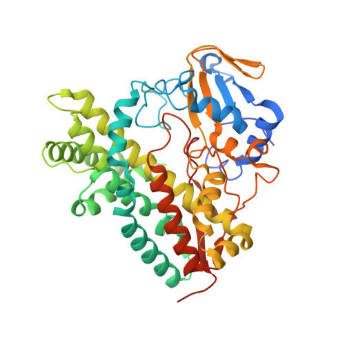
    CYP130 is one of the 20 Mycobacterium tuberculosis cytochrome P450 enzymes, only two of which, CYP51 and CYP121, have so far been studied as individually expressed proteins. Here we characterize a third heterologously expressed M. tuberculosis cytochrome P450, CYP130, by UV-visible spectroscopy, isothermal titration calorimetry, and x-ray crystallography, including determination of the crystal structures of ligand-free and econazole-bound CYP130 at a resolution of 1.46 and 3.0A(,) respectively. Ligand-free CYP130 crystallizes in an "open" conformation as a monomer, whereas the econazole-bound form crystallizes in a "closed" conformation as a dimer. Conformational changes enabling the "open-closed" transition involve repositioning of the BC-loop and the F and G helices that envelop the inhibitor in the binding site and reshape the protein surface. Crystal structure analysis shows that the portion of the BC-loop relocates as much as 18A between the open and closed conformations. Binding of econazole to CYP130 involves a conformational change and is mediated by both a set of hydrophobic interactions with amino acid residues in the active site and coordination of the heme iron. CYP130 also binds miconazole with virtually the same binding affinity as econazole and clotrimazole and ketoconazole with somewhat lower affinities, which makes it a plausible target for this class of therapeutic drugs. Overall, binding of the azole inhibitors is a sequential two-step, entropy-driven endothermic process. Binding of econazole and clotrimazole exhibits positive cooperativity that may reflect a propensity of CYP130 to associate into a dimeric structure.


  • Organizational Affiliation

    Department of Pharmaceutical Chemistry, University of California, San Francisco, California 94158, USA.


Macromolecules
Find similar proteins by:  (by identity cutoff)  |  3D Structure
Entity ID: 1
MoleculeChains Sequence LengthOrganismDetailsImage
CYTOCHROME P450 130
A, B
414Mycobacterium tuberculosis H37RvMutation(s): 0 
EC: 1.14
UniProt
Find proteins for P9WPN5 (Mycobacterium tuberculosis (strain ATCC 25618 / H37Rv))
Explore P9WPN5 
Go to UniProtKB:  P9WPN5
Entity Groups  
Sequence Clusters30% Identity50% Identity70% Identity90% Identity95% Identity100% Identity
UniProt GroupP9WPN5
Sequence Annotations
Expand
  • Reference Sequence
Small Molecules
Ligands 4 Unique
IDChains Name / Formula / InChI Key2D Diagram3D Interactions
HEM
Query on HEM

Download Ideal Coordinates CCD File 
C [auth A],
L [auth B]
PROTOPORPHYRIN IX CONTAINING FE
C34 H32 Fe N4 O4
KABFMIBPWCXCRK-RGGAHWMASA-L
ECN
Query on ECN

Download Ideal Coordinates CCD File 
D [auth A],
M [auth B]
1-[(2S)-2-[(4-CHLOROBENZYL)OXY]-2-(2,4-DICHLOROPHENYL)ETHYL]-1H-IMIDAZOLE
C18 H15 Cl3 N2 O
LEZWWPYKPKIXLL-GOSISDBHSA-N
SO4
Query on SO4

Download Ideal Coordinates CCD File 
H [auth A],
I [auth A],
J [auth A],
W [auth B]
SULFATE ION
O4 S
QAOWNCQODCNURD-UHFFFAOYSA-L
F
Query on F

Download Ideal Coordinates CCD File 
E [auth A]
F [auth A]
G [auth A]
K [auth A]
N [auth B]
E [auth A],
F [auth A],
G [auth A],
K [auth A],
N [auth B],
O [auth B],
P [auth B],
Q [auth B],
R [auth B],
S [auth B],
T [auth B],
U [auth B],
V [auth B],
X [auth B]
FLUORIDE ION
F
KRHYYFGTRYWZRS-UHFFFAOYSA-M
Experimental Data & Validation

Experimental Data

  • Method: X-RAY DIFFRACTION
  • Resolution: 3.00 Å
  • R-Value Free: 0.234 
  • R-Value Work: 0.200 
  • R-Value Observed: 0.200 
  • Space Group: P 32 2 1
Unit Cell:
Length ( Å )Angle ( ˚ )
a = 130.676α = 90
b = 130.676β = 90
c = 229.357γ = 120
Software Package:
Software NamePurpose
CNSrefinement
HKL-2000data reduction
SCALEPACKdata scaling
MOLREPphasing

Structure Validation

View Full Validation Report


Ligand Structure Quality Assessment 


Entry History 

Deposition Data

Revision History  (Full details and data files)

  • Version 1.0: 2007-12-18
    Type: Initial release
  • Version 1.1: 2011-08-17
    Changes: Atomic model, Version format compliance
  • Version 1.2: 2012-08-01
    Changes: Non-polymer description, Refinement description
  • Version 1.3: 2018-02-07
    Changes: Database references
  • Version 1.4: 2023-12-13
    Changes: Data collection, Database references, Derived calculations, Other, Refinement description