2UUK

Thrombin-hirugen-gw420128 ternary complex at 1.39A resolution


Experimental Data Snapshot

  • Method: X-RAY DIFFRACTION
  • Resolution: 1.39 Å
  • R-Value Free: 0.173 
  • R-Value Observed: 0.118 

wwPDB Validation   3D Report Full Report


This is version 1.6 of the entry. See complete history


Literature

The Determination of Protonation States in Proteins.

Ahmed, H.U.Blakeley, M.P.Cianci, M.Cruickshank, D.W.J.Hubbard, J.A.Helliwell, J.R.

(2007) Acta Crystallogr D Biol Crystallogr 63: 906

  • DOI: https://doi.org/10.1107/S0907444907029976
  • Primary Citation of Related Structures:  
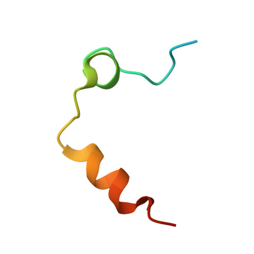
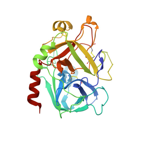
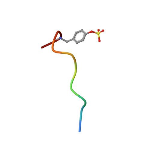
    2UU8, 2UUF, 2UUJ, 2UUK, 2YZ4

  • PubMed Abstract: 

    The protonation states of aspartic acids and glutamic acids as well as histidine are investigated in four X-ray cases: Ni,Ca concanavalin A at 0.94 A, a thrombin-hirugen binary complex at 1.26 A resolution and two thrombin-hirugen-inhibitor ternary complexes at 1.32 and 1.39 A resolution. The truncation of the Ni,Ca concanavalin A data at various test resolutions between 0.94 and 1.50 A provided a test comparator for the ;unknown' thrombin-hirugen carboxylate bond lengths. The protonation states of aspartic acids and glutamic acids can be determined (on the basis of convincing evidence) even to the modest resolution of 1.20 A as exemplified by our X-ray crystal structure refinements of Ni and Mn concanavalin A and also as indicated in the 1.26 A structure of thrombin, both of which are reported here. The protonation-state indication of an Asp or a Glu is valid provided that the following criteria are met (in order of importance). (i) The acidic residue must have a single occupancy. (ii) Anisotropic refinement at a minimum diffraction resolution of 1.20 A (X-ray data-to-parameter ratio of approximately 3.5:1) is required. (iii) Both of the bond lengths must agree with the expectation (i.e. dictionary values), thus allowing some relaxation of the bond-distance standard uncertainties required to approximately 0.025 A for a '3sigma' determination or approximately 0.04 A for a '2sigma' determination, although some variation of the expected bond-distance values must be allowed according to the microenvironment of the hydrogen of interest. (iv) Although the F(o) - F(c) map peaks are most likely to be unreliable at the resolution range around 1.20 A, if admitted as evidence the peak at the hydrogen position must be greater than or equal to 2.5 sigma and in the correct geometry. (v) The atomic B factors need to be less than 10 A(2) for bond-length differentiation; furthermore, the C=O bond can also be expected to be observed with continuous 2F(o) - F(c) electron density and the C-OH bond with discontinuous electron density provided that the atomic B factors are less than approximately 20 A(2) and the contour level is increased. The final decisive option is to carry out more than one experiment, e.g. multiple X-ray crystallography experiments and ideally neutron crystallography. The complementary technique of neutron protein crystallography has provided evidence of the protonation states of histidine and acidic residues in concanavalin A and also the correct orientations of asparagine and glutamine side chains. Again, the truncation of the neutron data at various test resolutions between 2.5 and 3.0 A, even 3.25 and 3.75 A resolution, examines the limits of the neutron probe. These various studies indicate a widening of the scope of both X-ray and neutron probes in certain circumstances to elucidate the protonation states in proteins.


  • Organizational Affiliation

    School of Chemistry, Brunswick Street, The University of Manchester, Manchester, England.


Macromolecules
Find similar proteins by:  (by identity cutoff)  |  3D Structure
Entity ID: 1
MoleculeChains Sequence LengthOrganismDetailsImage
HUMAN ALPHA THROMBIN36Homo sapiensMutation(s): 0 
EC: 3.4.21.5
UniProt & NIH Common Fund Data Resources
Find proteins for P00734 (Homo sapiens)
Explore P00734 
Go to UniProtKB:  P00734
PHAROS:  P00734
GTEx:  ENSG00000180210 
Entity Groups  
Sequence Clusters30% Identity50% Identity70% Identity90% Identity95% Identity100% Identity
UniProt GroupP00734
Sequence Annotations
Expand
  • Reference Sequence
Find similar proteins by:  (by identity cutoff)  |  3D Structure
Entity ID: 2
MoleculeChains Sequence LengthOrganismDetailsImage
THROMBIN259Homo sapiensMutation(s): 0 
EC: 3.4.21.5
UniProt & NIH Common Fund Data Resources
Find proteins for P00734 (Homo sapiens)
Explore P00734 
Go to UniProtKB:  P00734
PHAROS:  P00734
GTEx:  ENSG00000180210 
Entity Groups  
Sequence Clusters30% Identity50% Identity70% Identity90% Identity95% Identity100% Identity
UniProt GroupP00734
Sequence Annotations
Expand
  • Reference Sequence

Find similar proteins by:  Sequence   |   3D Structure  

Entity ID: 3
MoleculeChains Sequence LengthOrganismDetailsImage
HIRUDIN IC [auth H]10Hirudo medicinalisMutation(s): 0 
UniProt
Find proteins for P01050 (Hirudo medicinalis)
Explore P01050 
Go to UniProtKB:  P01050
Entity Groups  
Sequence Clusters30% Identity50% Identity70% Identity90% Identity95% Identity100% Identity
UniProt GroupP01050
Sequence Annotations
Expand
  • Reference Sequence
Small Molecules
Ligands 3 Unique
IDChains Name / Formula / InChI Key2D Diagram3D Interactions
897
Query on 897

Download Ideal Coordinates CCD File 
F [auth B]N-[3-(TERT-BUTYLAMINO)-3-OXOPROPYL]-N-ISOPROPYL-3-METHYL-5-{[(2S)-2-(PYRIDIN-4-YLAMINO)PROPYL]OXY}BENZAMIDE
C26 H38 N4 O3
VAFVRMZHAGPYIT-FQEVSTJZSA-N
CA
Query on CA

Download Ideal Coordinates CCD File 
E [auth B]CALCIUM ION
Ca
BHPQYMZQTOCNFJ-UHFFFAOYSA-N
NA
Query on NA

Download Ideal Coordinates CCD File 
D [auth B]SODIUM ION
Na
FKNQFGJONOIPTF-UHFFFAOYSA-N
Modified Residues  1 Unique
IDChains TypeFormula2D DiagramParent
TYS
Query on TYS
C [auth H]L-PEPTIDE LINKINGC9 H11 N O6 STYR
Experimental Data & Validation

Experimental Data

  • Method: X-RAY DIFFRACTION
  • Resolution: 1.39 Å
  • R-Value Free: 0.173 
  • R-Value Observed: 0.118 
  • Space Group: C 1 2 1
Unit Cell:
Length ( Å )Angle ( ˚ )
a = 70.346α = 90
b = 71.436β = 100.03
c = 72.116γ = 90
Software Package:
Software NamePurpose
DENZOdata reduction
SCALEPACKdata scaling
REFMACphasing
SHELXL-97phasing
SHELXL-97refinement

Structure Validation

View Full Validation Report


Entry History 

Deposition Data

Revision History  (Full details and data files)

  • Version 1.0: 2007-09-04
    Type: Initial release
  • Version 1.1: 2011-07-13
    Changes: Atomic model, Database references, Derived calculations, Non-polymer description, Structure summary, Version format compliance
  • Version 1.2: 2012-11-30
    Changes: Other
  • Version 1.3: 2013-03-13
    Changes: Other
  • Version 1.4: 2013-08-07
    Changes: Atomic model, Other
  • Version 1.5: 2019-05-22
    Changes: Data collection, Derived calculations, Other, Refinement description
  • Version 1.6: 2023-12-13
    Changes: Data collection, Database references, Derived calculations, Other, Refinement description