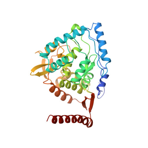
Crystal structure of tyrosine hydroxylase with bound cofactor analogue and iron at 2.3 A resolution: self-hydroxylation of Phe300 and the pterin-binding site.
Goodwill, K.E., Sabatier, C., Stevens, R.C.(1998) Biochemistry 37: 13437-13445
- PubMed: 9753429
- DOI: https://doi.org/10.1021/bi981462g
- Primary Citation of Related Structures:
2TOH - PubMed Abstract:
TyrOH is a non-heme iron enzyme which uses molecular oxygen to hydroxylate tyrosine to form L-dihydroxyphenylalanine (L-DOPA), and tetrahydrobiopterin to form 4a-hydroxybiopterin, in the rate-limiting step of the catecholamine biosynthetic pathway. The 2.3 A crystal structure of the catalytic and tetramerization domains of rat tyrosine hydroxylase (TyrOH) in the presence of the cofactor analogue 7,8-dihydrobiopterin and iron shows the mode of pterin binding and the proximity of its hydroxylated 4a carbon to the required iron. The pterin binds on one face of the large active-site cleft, forming an aromatic pi-stacking interaction with Phe300. This phenylalanine residue of TyrOH is found to be hydroxylated in the meta position, most likely through an autocatalytic process, and to consequently form a hydrogen bond to the main-chain carbonyl of Gln310 which anchors Phe300 in the active site. The bound pterin forms hydrogen bonds from N-8 to the main-chain carbonyl of Leu295, from O-4 to Tyr371 and Glu376, from the C-1' OH to the main-chain amides of Leu294 and Leu295, and from the C-2' hydroxyl to an iron-coordinating water. The part of the pterin closest to the iron is the O-4 carbonyl oxygen at a distance of 3.6 A. The iron is 5.6 A from the pterin 4a carbon which is hydroxylated in the enzymatic reaction. No structural changes are observed between the pterin bound and the nonliganded enzyme. On the basis of these structures, molecular oxygen could bind in a bridging position optimally between the pterin C-4a and iron atom prior to substrate hydroxylation. This structure represents the first report of close interactions between pterin and iron in an enzyme active site.
Organizational Affiliation:
Department of Chemistry, University of California, Berkeley, California 94720, USA.