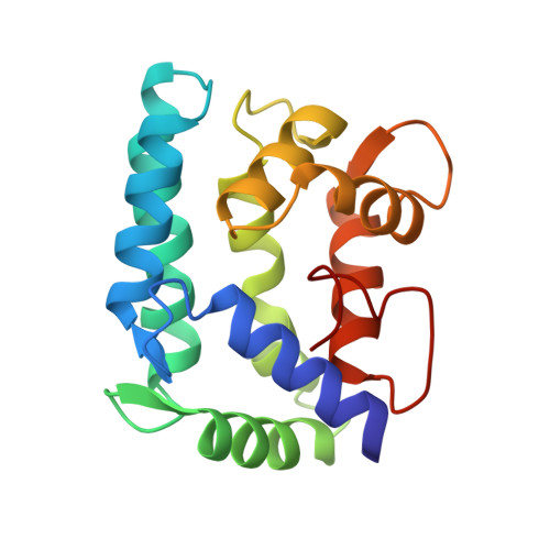
Structure of a sarcoplasmic calcium-binding protein from Nereis diversicolor refined at 2.0 A resolution.
Vijay-Kumar, S., Cook, W.J.(1992) J Mol Biol 224: 413-426
- PubMed: 1560459
- DOI: https://doi.org/10.1016/0022-2836(92)91004-9
- Primary Citation of Related Structures:
2SCP - PubMed Abstract:
The crystal structure of a sarcoplasmic Ca(2+)-binding protein (SCP) from the sandworm Nereis diversicolor has been determined and refined at 2.0 A resolution using restrained least-squares techniques. The two molecules in the crystallographic asymmetric unit, which are related by a non-crystallographic 2-fold axis, were refined independently. The refined model includes all 174 residues and three calcium ions for each molecule, as well as 213 water molecules. The root-mean-square difference in co-ordinates for backbone atoms and calcium ions of the two molecules is 0.51 A. The final crystallographic R-factor, based on 18,959 reflections in the range 2.0 A less than or equal to d less than or equal to 7.0 A, with intensities exceeding 2.0 sigma, is 0.182. Bond lengths and bond angles in the molecules have root-mean-square deviations from ideal values of 0.013 A and 2.2 degrees, respectively. SCP has four distinct domains with the typical helix-loop-helix (EF-hand) Ca(2+)-binding motif, although the second Ca(2+)-binding domain is not functional due to amino acid changes in the loop. The structure shows several unique features compared to other Ca(2+)-binding proteins with four EF-hand domains. The overall structure is highly compact and globular with a predominant hydrophobic core, unlike the extended dumbbell-shaped structure of calmodulin or troponin C. A hydrophobic tail at the COOH terminus adds to the structural stability by packing against a hydrophobic pocket created by the folding of the NH2 and COOH-terminal Ca(2+)-binding domain pairs. The first and second domains show different helix-packing arrangements from any previously described for Ca(2+)-binding proteins.
Organizational Affiliation:
Department of Biochemistry, University of Alabama, Birmingham.