2ROK

Solution structure of the cap-binding domain of PARN complexed with the cap analog


Experimental Data Snapshot

  • Method: SOLUTION NMR
  • Conformers Calculated: 40 
  • Conformers Submitted: 20 
  • Selection Criteria: structures with the lowest energy 

wwPDB Validation   3D Report Full Report


This is version 1.2 of the entry. See complete history


Literature

The RRM domain of poly(A)-specific ribonuclease has a noncanonical binding site for mRNA cap analog recognition.

Nagata, T.Suzuki, S.Endo, R.Shirouzu, M.Terada, T.Inoue, M.Kigawa, T.Kobayashi, N.Guntert, P.Tanaka, A.Hayashizaki, Y.Muto, Y.Yokoyama, S.

(2008) Nucleic Acids Res 36: 4754-4767

  • DOI: https://doi.org/10.1093/nar/gkn458
  • Primary Citation of Related Structures:  
    2ROK

  • PubMed Abstract: 

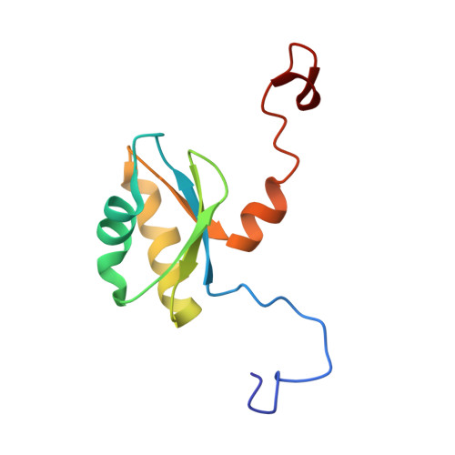
    The degradation of the poly(A) tail is crucial for posttranscriptional gene regulation and for quality control of mRNA. Poly(A)-specific ribonuclease (PARN) is one of the major mammalian 3' specific exo-ribonucleases involved in the degradation of the mRNA poly(A) tail, and it is also involved in the regulation of translation in early embryonic development. The interaction between PARN and the m(7)GpppG cap of mRNA plays a key role in stimulating the rate of deadenylation. Here we report the solution structures of the cap-binding domain of mouse PARN with and without the m(7)GpppG cap analog. The structure of the cap-binding domain adopts the RNA recognition motif (RRM) with a characteristic alpha-helical extension at its C-terminus, which covers the beta-sheet surface (hereafter referred to as PARN RRM). In the complex structure of PARN RRM with the cap analog, the base of the N(7)-methyl guanosine (m(7)G) of the cap analog stacks with the solvent-exposed aromatic side chain of the distinctive tryptophan residue 468, located at the C-terminal end of the second beta-strand. These unique structural features in PARN RRM reveal a novel cap-binding mode, which is distinct from the nucleotide recognition mode of the canonical RRM domains.


  • Organizational Affiliation

    Systems and Structural Biology Center, Yokohama Institute, RIKEN, 1-7-22 Suehiro-cho, Tsurumi-ku, Yokohama, Japan.


Macromolecules
Find similar proteins by:  (by identity cutoff)  |  3D Structure
Entity ID: 1
MoleculeChains Sequence LengthOrganismDetailsImage
poly(A)-specific ribonuclease100Mus musculusMutation(s): 0 
Gene Names: Parn
EC: 3.1.13.4
UniProt
Find proteins for Q8VDG3 (Mus musculus)
Explore Q8VDG3 
Go to UniProtKB:  Q8VDG3
Entity Groups  
Sequence Clusters30% Identity50% Identity70% Identity90% Identity95% Identity100% Identity
UniProt GroupQ8VDG3
Sequence Annotations
Expand
  • Reference Sequence
Small Molecules
Ligands 2 Unique
IDChains Name / Formula / InChI Key2D Diagram3D Interactions
GDP
Query on GDP

Download Ideal Coordinates CCD File 
C [auth A]GUANOSINE-5'-DIPHOSPHATE
C10 H15 N5 O11 P2
QGWNDRXFNXRZMB-UUOKFMHZSA-N
7MG
Query on 7MG

Download Ideal Coordinates CCD File 
B [auth A]7N-METHYL-8-HYDROGUANOSINE-5'-MONOPHOSPHATE
C11 H18 N5 O8 P
ZMWJGXGSWZFZPJ-KQYNXXCUSA-N
Binding Affinity Annotations 
IDSourceBinding Affinity
7MG PDBBind:  2ROK Kd: 4.50e+4 (nM) from 1 assay(s)
Experimental Data & Validation

Experimental Data

  • Method: SOLUTION NMR
  • Conformers Calculated: 40 
  • Conformers Submitted: 20 
  • Selection Criteria: structures with the lowest energy 

Structure Validation

View Full Validation Report


Entry History 

Revision History  (Full details and data files)

  • Version 1.0: 2009-02-10
    Type: Initial release
  • Version 1.1: 2011-07-13
    Changes: Version format compliance
  • Version 1.2: 2022-03-16
    Changes: Data collection, Database references, Derived calculations