Solution structure of the DNA binding domain of rice telomere binding protein RTBP1
Ko, S., Yu, E.Y., Shin, J., Yoo, H.H., Tanaka, T., Kim, W.T., Cho, H.S., Lee, W., Chung, I.K.(2009) Biochemistry 48: 827-838
- PubMed: 19152316
- DOI: https://doi.org/10.1021/bi801270g
- Primary Citation of Related Structures:
2ROH - PubMed Abstract:
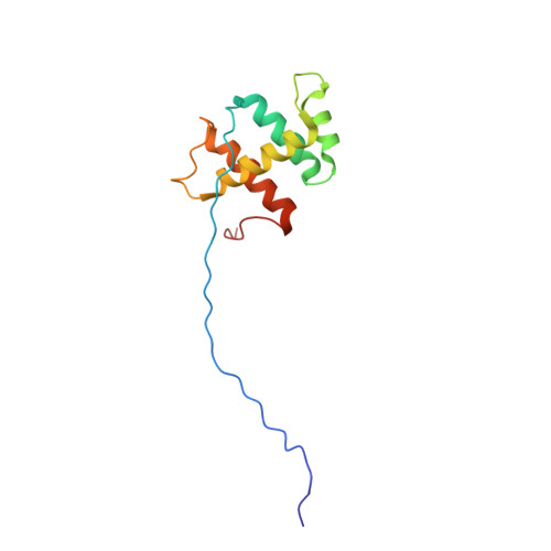
RTBP1 is a rice telomeric protein that binds to the duplex array of TTTAGGG repeats at chromosome ends. The DNA binding domain of RTBP1 contains a Myb-type DNA binding motif and a highly conserved C-terminal Myb extension that is unique to plant telomeric proteins. Using an electrophoretic mobility shift assay, we identified the C-terminal 110-amino acid region (RTBP1(506-615)) as the minimal telomeric DNA binding domain, suggesting that the Myb extension is required for binding plant telomeric DNA. Like other telomeric proteins such as human TRF1 and yeast Rap1, RTBP1 induced a DNA bending in the telomeric repeat sequence, suggesting that RTBP1 may play a role in establishing and/or maintaining an active telomere configuration in vivo. To elucidate the DNA binding mode of RTBP1, we determined the three-dimensional structure of RTBP1(506-615) in solution by NMR spectroscopy. The overall structure of RTBP1(506-615) is composed of four alpha-helices and stabilized by three hydrophobic patches. The second and third helices in RTBP1 form a helix-turn-helix motif that interacts directly with DNA. The fourth helix located in the Myb extension is essential for binding to telomeric DNA via stabilization of the overall structure of the RTBP1 DNA binding domain. When DNA bound to RTBP1(506-615), large chemical shift perturbations were induced in the N-terminal arm, helix 3, and the loop between helices 3 and 4. These results suggest that helix 3 functions as a sequence-specific recognition helix while the N-terminal arm stabilizes the DNA binding.
Organizational Affiliation:
Department of Biochemistry and Biology, Protein Network Research Center, Yonsei University, Seoul 120-749, Korea.