Common and divergent structural features of a series of corticotropin releasing factor-related peptides
Grace, C.R.R., Perrin, M.H., Cantle, J.P., Vale, W.W., Rivier, J.E., Riek, R.(2007) J Am Chem Soc 129: 16102-16114
- PubMed: 18052377
- DOI: https://doi.org/10.1021/ja0760933
- Primary Citation of Related Structures:
2RM9, 2RMD, 2RME, 2RMF, 2RMG, 2RMH - PubMed Abstract:
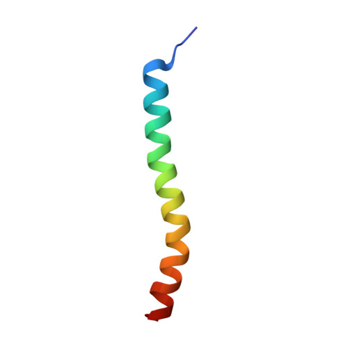
Members of the corticoliberin family include the corticotropin releasing factors (CRFs), sauvagine, the urotensins, and urocortin 1 (Ucn1), which bind to both the CRF receptors CRF-R1 and CRF-R2, and the urocortins 2 (Ucn2) and 3 (Ucn3), which are selective agonists of CRF-R2. Structure activity relationship studies led to several potent and long-acting analogues with selective binding to either one of the receptors. NMR structures of six ligands of this family (the antagonists astressin B and astressin2-B, the agonists stressin1, and the natural ligands human Ucn1, Ucn2, and Ucn3) were determined in DMSO. These six peptides show differences in binding affinities, receptor-selectivity, and NMR structure. Overall, their backbones are alpha-helical, with a small kink or a turn around residues 25-27, resulting in a helix-loop-helix motif. The C-terminal helices are of amphipathic nature, whereas the N-terminal helices vary in their amphipathicity. The C-terminal helices thereby assume a conformation very similar to that of astressin bound to the ECD1 of CRF-R2 recently reported by our group.1 On the basis of an analysis of the observed 3D structures and relative potencies of [Ala]-substituted analogues, it is proposed that both helices could play a crucial role in receptor binding and selectivity. In conclusion, the C-terminal helices may interact along their hydrophobic faces with the ECD1, whereas the entire N-terminal helical surface may be involved in receptor activation. On the basis of the common and divergent features observed in the 3D structures of these ligands, multiple binding models are proposed that may explain their plurality of actions.
Organizational Affiliation:
Structural Biology Laboratory, The Salk Institute for Biological Studies, 10010 North Torrey Pines Road, La Jolla, California 92037, USA.