2RK8

The Structure of rat cytosolic PEPCK in complex with phosphonoformate


Experimental Data Snapshot

  • Method: X-RAY DIFFRACTION
  • Resolution: 2.00 Å
  • R-Value Free: 0.249 
  • R-Value Work: 0.195 
  • R-Value Observed: 0.198 

wwPDB Validation   3D Report Full Report


This is version 1.3 of the entry. See complete history


Literature

Differential Inhibition of Cytosolic PEPCK by Substrate Analogues. Kinetic and Structural Characterization of Inhibitor Recognition.

Stiffin, R.M.Sullivan, S.M.Carlson, G.M.Holyoak, T.

(2008) Biochemistry 47: 2099-2109

  • DOI: https://doi.org/10.1021/bi7020662
  • Primary Citation of Related Structures:  
    2RK7, 2RK8, 2RKA, 2RKD, 2RKE

  • PubMed Abstract: 

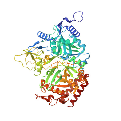
    The mechanisms of molecular recognition of phosphoenolpyruvate (PEP) and oxaloacetate (OAA) by cytosolic phosphoenolpyruvate carboxykinase (cPEPCK) were investigated by the systematic evaluation of a variety of PEP and OAA analogues as potential reversible inhibitors of the enzyme against PEP. The molecules that inhibit the enzyme in a competitive fashion were found to fall into two general classes. Those molecules that mimic the binding geometry of PEP, namely phosphoglycolate and 3-phosphonopropionate, are found to bind weakly (millimolar Ki values). In contrast, those competitive inhibitors that mimic the binding of OAA (oxalate and phosphonoformate) coordinate directly to the active site manganese ion and bind an order of magnitude more tightly (micromolar Ki values). The competitive inhibitor sulfoacetate is found to be an outlier of these two classes, binding in a hybrid fashion utilizing modes of recognition of both PEP and OAA in order to achieve a micromolar inhibition constant in the absence of direct coordination to the active site metal. The kinetic studies in combination with the structural characterization of the five aforementioned competitive inhibitors demonstrate the molecular requirements for high affinity binding of molecules to the active site of the enzyme. These features include cis-planar carbonyl groups that are required for coordination to the active site metal, a bridging electron rich atom at the position corresponding to the C2 methylene group of OAA to facilitate interactions with R405, a carboxylate or sulfonate moiety at a position corresponding to the C1 carboxylate of OAA, and the edge-on aromatic interaction between a carboxylate and Y235.


  • Organizational Affiliation

    Department of Molecular Biosciences, The University of Tennessee Health Science Center, Memphis, Tennessee 38163, USA.


Macromolecules
Find similar proteins by:  (by identity cutoff)  |  3D Structure
Entity ID: 1
MoleculeChains Sequence LengthOrganismDetailsImage
Phosphoenolpyruvate carboxykinase, cytosolic [GTP]
A, B
624Rattus norvegicusMutation(s): 0 
Gene Names: Pck1
EC: 4.1.1.32
UniProt
Find proteins for P07379 (Rattus norvegicus)
Explore P07379 
Go to UniProtKB:  P07379
Entity Groups  
Sequence Clusters30% Identity50% Identity70% Identity90% Identity95% Identity100% Identity
UniProt GroupP07379
Sequence Annotations
Expand
  • Reference Sequence
Small Molecules
Ligands 5 Unique
IDChains Name / Formula / InChI Key2D Diagram3D Interactions
PPF
Query on PPF

Download Ideal Coordinates CCD File 
E [auth A],
J [auth B]
PHOSPHONOFORMIC ACID
C H3 O5 P
ZJAOAACCNHFJAH-UHFFFAOYSA-N
PEG
Query on PEG

Download Ideal Coordinates CCD File 
G [auth A]DI(HYDROXYETHYL)ETHER
C4 H10 O3
MTHSVFCYNBDYFN-UHFFFAOYSA-N
MN
Query on MN

Download Ideal Coordinates CCD File 
C [auth A],
H [auth B]
MANGANESE (II) ION
Mn
WAEMQWOKJMHJLA-UHFFFAOYSA-N
FMT
Query on FMT

Download Ideal Coordinates CCD File 
F [auth A],
K [auth B]
FORMIC ACID
C H2 O2
BDAGIHXWWSANSR-UHFFFAOYSA-N
NA
Query on NA

Download Ideal Coordinates CCD File 
D [auth A],
I [auth B]
SODIUM ION
Na
FKNQFGJONOIPTF-UHFFFAOYSA-N
Binding Affinity Annotations 
IDSourceBinding Affinity
PPF PDBBind:  2RK8 Ki: 2.30e+5 (nM) from 1 assay(s)
Binding MOAD:  2RK8 Ki: 2.30e+5 (nM) from 1 assay(s)
Experimental Data & Validation

Experimental Data

  • Method: X-RAY DIFFRACTION
  • Resolution: 2.00 Å
  • R-Value Free: 0.249 
  • R-Value Work: 0.195 
  • R-Value Observed: 0.198 
  • Space Group: P 1 21 1
Unit Cell:
Length ( Å )Angle ( ˚ )
a = 62.251α = 90
b = 119.47β = 107.06
c = 86.87γ = 90
Software Package:
Software NamePurpose
DENZOdata reduction
SCALEPACKdata scaling
REFMACrefinement
PDB_EXTRACTdata extraction
CrystalCleardata collection

Structure Validation

View Full Validation Report


Entry History 

Deposition Data

Revision History  (Full details and data files)

  • Version 1.0: 2008-01-29
    Type: Initial release
  • Version 1.1: 2011-07-13
    Changes: Advisory, Version format compliance
  • Version 1.2: 2017-10-25
    Changes: Refinement description
  • Version 1.3: 2024-02-21
    Changes: Data collection, Database references, Derived calculations