2RGW

Catalytic Subunit of M. jannaschii Aspartate Transcarbamoylase


Experimental Data Snapshot

  • Method: X-RAY DIFFRACTION
  • Resolution: 2.80 Å
  • R-Value Free: 0.257 
  • R-Value Work: 0.199 

wwPDB Validation   3D Report Full Report


This is version 1.2 of the entry. See complete history


Literature

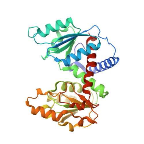
Crystal structure of the catalytic trimer of Methanococcus jannaschii aspartate transcarbamoylase.

Vitali, J.Colaneri, M.J.Kantrowitz, E.

(2008) Proteins 71: 1324-1334

  • DOI: https://doi.org/10.1002/prot.21667
  • Primary Citation of Related Structures:  
    2RGW

  • PubMed Abstract: 

    The catalytic trimer of Methanococcus jannaschii aspartate transcarbamoylase is extremely heat stable, maintaining 75% of its activity after heat treatment for 60 min at 75 degrees C. We undertook its structural analysis in order to understand the molecular basis of its thermostability and gain insight on how its catalytic function adapts to high temperature. Several structural elements potentially contributing to thermostability were identified. These include: (i) changes in the amino acid composition such as a decrease in the thermolabile residues Gln and Asn, an increase in the charged residues Lys and Glu, an increase in Tyr and a decrease in Ala residues; (ii) a larger number of salt bridges, in particular, the improvement of ion-pair networks; (iii) shortening of the N-terminus and shortening of three loops. An interesting feature of the crystal structure is the association of two crystallographically independent catalytic subunits into a staggered complex with an intertrimer distance of 33.8 A. The active site appears similar to Escherichia coli. Upon substrate binding, smaller changes in the global orientation of domains and larger conformational changes of the active site residues are expected as compared to E. coli.


  • Organizational Affiliation

    Department of Physics, Cleveland State University, Euclid Avenue at East 24th Street, Cleveland, Ohio 44115, USA. j.vitali@csuohio.edu


Macromolecules
Find similar proteins by:  (by identity cutoff)  |  3D Structure
Entity ID: 1
MoleculeChains Sequence LengthOrganismDetailsImage
Aspartate carbamoyltransferase
A, B, C, D, E
A, B, C, D, E, F
306N/AMutation(s): 0 
Gene Names: pyrB
EC: 2.1.3.2
UniProt
Find proteins for Q58976 (Methanocaldococcus jannaschii (strain ATCC 43067 / DSM 2661 / JAL-1 / JCM 10045 / NBRC 100440))
Explore Q58976 
Go to UniProtKB:  Q58976
Entity Groups  
Sequence Clusters30% Identity50% Identity70% Identity90% Identity95% Identity100% Identity
UniProt GroupQ58976
Sequence Annotations
Expand
  • Reference Sequence
Small Molecules
Ligands 1 Unique
IDChains Name / Formula / InChI Key2D Diagram3D Interactions
SO4
Query on SO4

Download Ideal Coordinates CCD File 
G [auth A],
H [auth D]
SULFATE ION
O4 S
QAOWNCQODCNURD-UHFFFAOYSA-L
Experimental Data & Validation

Experimental Data

  • Method: X-RAY DIFFRACTION
  • Resolution: 2.80 Å
  • R-Value Free: 0.257 
  • R-Value Work: 0.199 
  • Space Group: C 1 2 1
Unit Cell:
Length ( Å )Angle ( ˚ )
a = 139.9α = 90
b = 91.89β = 98.3
c = 154.2γ = 90
Software Package:
Software NamePurpose
EPMRphasing
CNSrefinement
DENZOdata reduction
SCALEPACKdata scaling

Structure Validation

View Full Validation Report


Entry History 

Deposition Data

Revision History  (Full details and data files)

  • Version 1.0: 2008-10-14
    Type: Initial release
  • Version 1.1: 2011-07-13
    Changes: Version format compliance
  • Version 1.2: 2023-08-30
    Changes: Data collection, Database references, Derived calculations, Refinement description