2RGV

The crystal structure of PerR-Ox highlights 2-oxo-Histidine formation


Experimental Data Snapshot

  • Method: X-RAY DIFFRACTION
  • Resolution: 2.05 Å
  • R-Value Free: 0.308 
  • R-Value Work: 0.211 
  • R-Value Observed: 0.216 

wwPDB Validation   3D Report Full Report


This is version 1.1 of the entry. See complete history


Literature

Structural and functional characterization of 2-oxo-histidine in oxidized PerR protein.

Traore, D.A.El Ghazouani, A.Jacquamet, L.Borel, F.Ferrer, J.L.Lascoux, D.Ravanat, J.L.Jaquinod, M.Blondin, G.Caux-Thang, C.Duarte, V.Latour, J.M.

(2009) Nat Chem Biol 5: 53-59

  • DOI: https://doi.org/10.1038/nchembio.133
  • Primary Citation of Related Structures:  
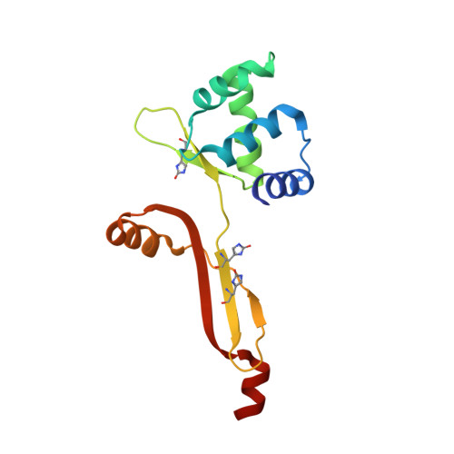
    2RGV

  • PubMed Abstract: 

    In Bacillus subtilis, PerR is a metal-dependent sensor of hydrogen peroxide. PerR is a dimeric zinc protein with a regulatory site that coordinates either Fe(2+) (PerR-Zn-Fe) or Mn(2+) (PerR-Zn-Mn). Though most of the peroxide sensors use cysteines to detect H(2)O(2), it has been shown that reaction of PerR-Zn-Fe with H(2)O(2) leads to the oxidation of one histidine residue. Oxidation of PerR leads to the incorporation of one oxygen atom into His37 or His91. This study presents the crystal structure of the oxidized PerR protein (PerR-Zn-ox), which clearly shows a 2-oxo-histidine residue in position 37. Formation of 2-oxo-histidine is demonstrated and quantified by HPLC-MS/MS. EPR experiments indicate that PerR-Zn-H37ox retains a significant affinity for the regulatory metal, whereas PerR-Zn-H91ox shows a considerably reduced affinity for the metal ion. In spite of these major differences in terms of metal binding affinity, oxidation of His37 and/or His91 in PerR prevents DNA binding.


  • Organizational Affiliation

    Commissariat à l'Energie Atomique, Institut de Recherches en Technologies et Sciences pour le Vivant, Laboratoire de Chimie et Biologie des Métaux, CEA-Grenoble, 17 avenue des Martyrs, 38054 Grenoble Cedex 9, France.


Macromolecules
Find similar proteins by:  (by identity cutoff)  |  3D Structure
Entity ID: 1
MoleculeChains Sequence LengthOrganismDetailsImage
Peroxide operon regulator
A, B
145Bacillus subtilisMutation(s): 0 
UniProt
Find proteins for P71086 (Bacillus subtilis (strain 168))
Explore P71086 
Go to UniProtKB:  P71086
Entity Groups  
Sequence Clusters30% Identity50% Identity70% Identity90% Identity95% Identity100% Identity
UniProt GroupP71086
Sequence Annotations
Expand
  • Reference Sequence
Small Molecules
Modified Residues  1 Unique
IDChains TypeFormula2D DiagramParent
OHI
Query on OHI
A, B
L-PEPTIDE LINKINGC6 H7 N3 O3HIS
Experimental Data & Validation

Experimental Data

  • Method: X-RAY DIFFRACTION
  • Resolution: 2.05 Å
  • R-Value Free: 0.308 
  • R-Value Work: 0.211 
  • R-Value Observed: 0.216 
  • Space Group: P 1
Unit Cell:
Length ( Å )Angle ( ˚ )
a = 41.25α = 82.05
b = 42.7β = 79.98
c = 53.02γ = 61.63
Software Package:
Software NamePurpose
REFMACrefinement
PDB_EXTRACTdata extraction
ADSCdata collection
XDSdata reduction
XSCALEdata scaling
PHASERphasing

Structure Validation

View Full Validation Report


Entry History 

Deposition Data

Revision History  (Full details and data files)

  • Version 1.0: 2008-10-14
    Type: Initial release
  • Version 1.1: 2011-07-13
    Changes: Version format compliance