2RCT

Crystal structure of human holo cellular retinol-binding protein II (CRBP-II)


Experimental Data Snapshot

  • Method: X-RAY DIFFRACTION
  • Resolution: 1.20 Å
  • R-Value Free: 0.215 
  • R-Value Work: 0.193 
  • R-Value Observed: 0.194 

wwPDB Validation   3D Report Full Report


Ligand Structure Quality Assessment 


This is version 1.2 of the entry. See complete history


Literature

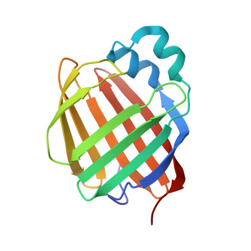
Crystal structure of human cellular retinol-binding protein II to 1.2 A resolution.

Tarter, M.Capaldi, S.Carrizo, M.E.Ambrosi, E.Perduca, M.Monaco, H.L.

(2007) Proteins 70: 1626-1630


Macromolecules
Find similar proteins by:  (by identity cutoff)  |  3D Structure
Entity ID: 1
MoleculeChains Sequence LengthOrganismDetailsImage
Retinol-binding protein II, cellular141Homo sapiensMutation(s): 0 
Gene Names: RBP2CRBP2
UniProt & NIH Common Fund Data Resources
Find proteins for P50120 (Homo sapiens)
Explore P50120 
Go to UniProtKB:  P50120
PHAROS:  P50120
GTEx:  ENSG00000114113 
Entity Groups  
Sequence Clusters30% Identity50% Identity70% Identity90% Identity95% Identity100% Identity
UniProt GroupP50120
Sequence Annotations
Expand
  • Reference Sequence
Experimental Data & Validation

Experimental Data

  • Method: X-RAY DIFFRACTION
  • Resolution: 1.20 Å
  • R-Value Free: 0.215 
  • R-Value Work: 0.193 
  • R-Value Observed: 0.194 
  • Space Group: P 21 21 21
Unit Cell:
Length ( Å )Angle ( ˚ )
a = 35.45α = 90
b = 49.966β = 90
c = 74.296γ = 90
Software Package:
Software NamePurpose
REFMACrefinement
ADSCdata collection
MOSFLMdata reduction
SCALAdata scaling
XFITdata reduction

Structure Validation

View Full Validation Report


Ligand Structure Quality Assessment 


Entry History 

Deposition Data

Revision History  (Full details and data files)

  • Version 1.0: 2007-10-02
    Type: Initial release
  • Version 1.1: 2011-07-13
    Changes: Version format compliance
  • Version 1.2: 2023-08-30
    Changes: Data collection, Database references, Derived calculations, Refinement description