2R83

Crystal structure analysis of human synaptotagmin 1 C2A-C2B


Experimental Data Snapshot

  • Method: X-RAY DIFFRACTION
  • Resolution: 2.70 Å
  • R-Value Free: 0.253 
  • R-Value Work: 0.230 
  • R-Value Observed: 0.230 

wwPDB Validation   3D Report Full Report


This is version 1.3 of the entry. See complete history


Literature

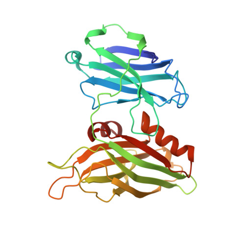
Structure of human synaptotagmin 1 C2AB in the absence of Ca2+ reveals a novel domain association.

Fuson, K.L.Montes, M.Robert, J.J.Sutton, R.B.

(2007) Biochemistry 46: 13041-13048

  • DOI: https://doi.org/10.1021/bi701651k
  • Primary Citation of Related Structures:  
    2R83

  • PubMed Abstract: 

    Release of neurotransmitter from synaptic vesicles requires the Ca2+/phospholipid-binding protein synaptotagmin 1. There is considerable evidence that cooperation between the tandem C2 domains of synaptotagmin is a requirement of regulated exocytosis; however, high-resolution structural evidence for this interaction has been lacking. The 2.7 A crystal structure of the cytosolic domains of human synaptotagmin 1 in the absence of Ca2+ reveals a novel closed conformation of the protein. The shared interface between C2A and C2B is stabilized by a network of interactions between residues on the C-terminal alpha-helix of the C2B domain and residues on loops 1-3 of the Ca2+-binding region of C2A. These interactions alter the overall shape of the Ca2+-binding pocket of C2A, but not that of C2B. Thus, synaptotagmin 1 C2A-C2B may utilize a novel regulatory mechanism whereby one C2 domain could regulate the other until an appropriate triggering event decouples them.


  • Organizational Affiliation

    Department of Biochemistry and Molecular Biology, The University of Texas Medical Branch, Galveston, Texas 77555, USA.


Macromolecules
Find similar proteins by:  (by identity cutoff)  |  3D Structure
Entity ID: 1
MoleculeChains Sequence LengthOrganismDetailsImage
Synaptotagmin-1
A, B
284Homo sapiensMutation(s): 0 
Gene Names: SYT1SVP65SYT
Membrane Entity: Yes 
UniProt & NIH Common Fund Data Resources
Find proteins for P21579 (Homo sapiens)
Explore P21579 
Go to UniProtKB:  P21579
PHAROS:  P21579
GTEx:  ENSG00000067715 
Entity Groups  
Sequence Clusters30% Identity50% Identity70% Identity90% Identity95% Identity100% Identity
UniProt GroupP21579
Sequence Annotations
Expand
  • Reference Sequence
Experimental Data & Validation

Experimental Data

  • Method: X-RAY DIFFRACTION
  • Resolution: 2.70 Å
  • R-Value Free: 0.253 
  • R-Value Work: 0.230 
  • R-Value Observed: 0.230 
  • Space Group: P 21 21 21
Unit Cell:
Length ( Å )Angle ( ˚ )
a = 82.372α = 90
b = 86.31β = 90
c = 147.186γ = 90
Software Package:
Software NamePurpose
PHASERphasing
CNSrefinement
ADSCdata collection
SCALAdata scaling

Structure Validation

View Full Validation Report


Entry History 

Deposition Data

Revision History  (Full details and data files)

  • Version 1.0: 2008-02-12
    Type: Initial release
  • Version 1.1: 2011-07-13
    Changes: Version format compliance
  • Version 1.2: 2018-01-24
    Changes: Structure summary
  • Version 1.3: 2023-08-30
    Changes: Data collection, Database references, Derived calculations, Refinement description