2R6N

Crystal structure of a pyrrolopyrimidine inhibitor in complex with human Cathepsin K


Experimental Data Snapshot

  • Method: X-RAY DIFFRACTION
  • Resolution: 1.95 Å
  • R-Value Free: 0.184 
  • R-Value Work: 0.156 

wwPDB Validation   3D Report Full Report


Ligand Structure Quality Assessment 


This is version 1.2 of the entry. See complete history


Literature

Novel scaffold for cathepsin K inhibitors.

Teno, N.Miyake, T.Ehara, T.Irie, O.Sakaki, J.Ohmori, O.Gunji, H.Matsuura, N.Masuya, K.Hitomi, Y.Nonomura, K.Horiuchi, M.Gohda, K.Iwasaki, A.Umemura, I.Tada, S.Kometani, M.Iwasaki, G.Cowan-Jacob, S.W.Missbach, M.Lattmann, R.Betschart, C.

(2007) Bioorg Med Chem Lett 17: 6096-6100

  • DOI: https://doi.org/10.1016/j.bmcl.2007.09.047
  • Primary Citation of Related Structures:  
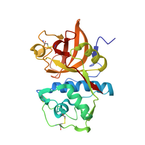
    2R6N

  • PubMed Abstract: 

    Pyrrolopyrimidine, a novel scaffold, allows to adjust interactions within the S3 subsite of cathepsin K. The core intermediate 10 facilitated the P3 optimization and identified highly potent and selective cathepsin K inhibitors 11-20.


  • Organizational Affiliation

    Novartis Institutes for BioMedical Research, Ohkubo 8, Tsukuba, Ibaraki, Japan. naoki.teno@novartis.com


Macromolecules
Find similar proteins by:  (by identity cutoff)  |  3D Structure
Entity ID: 1
MoleculeChains Sequence LengthOrganismDetailsImage
Cathepsin K217Homo sapiensMutation(s): 0 
Gene Names: CTSKCTSOCTSO2
EC: 3.4.22.38
UniProt & NIH Common Fund Data Resources
Find proteins for P43235 (Homo sapiens)
Explore P43235 
Go to UniProtKB:  P43235
PHAROS:  P43235
GTEx:  ENSG00000143387 
Entity Groups  
Sequence Clusters30% Identity50% Identity70% Identity90% Identity95% Identity100% Identity
UniProt GroupP43235
Sequence Annotations
Expand
  • Reference Sequence
Small Molecules
Ligands 1 Unique
IDChains Name / Formula / InChI Key2D Diagram3D Interactions
CKE
Query on CKE

Download Ideal Coordinates CCD File 
B [auth A]1-{7-cyclohexyl-6-[4-(4-methylpiperazin-1-yl)benzyl]-7H-pyrrolo[2,3-d]pyrimidin-2-yl}methanamine
C25 H34 N6
GCJSOJRPNOWSEH-UHFFFAOYSA-N
Binding Affinity Annotations 
IDSourceBinding Affinity
CKE PDBBind:  2R6N IC50: 1 (nM) from 1 assay(s)
Experimental Data & Validation

Experimental Data

  • Method: X-RAY DIFFRACTION
  • Resolution: 1.95 Å
  • R-Value Free: 0.184 
  • R-Value Work: 0.156 
  • Space Group: P 61
Unit Cell:
Length ( Å )Angle ( ˚ )
a = 62.4α = 90
b = 62.4β = 90
c = 121.8γ = 120
Software Package:
Software NamePurpose
AMoREphasing
CNSrefinement
PDB_EXTRACTdata extraction
MAR345dtbdata collection
XDSdata reduction
AUTOMARdata reduction

Structure Validation

View Full Validation Report


Ligand Structure Quality Assessment 


Entry History 

Deposition Data

Revision History  (Full details and data files)

  • Version 1.0: 2007-11-06
    Type: Initial release
  • Version 1.1: 2011-07-13
    Changes: Version format compliance
  • Version 1.2: 2017-10-25
    Changes: Advisory, Refinement description