The Structure of Sucrose Phosphate Synthase from Halothermothrix orenii Reveals Its Mechanism of Action and Binding Mode.
Chua, T.K., Bujnicki, J.M., Tan, T.C., Huynh, F., Patel, B.K., Sivaraman, J.(2008) Plant Cell 20: 1059-1072
- PubMed: 18424616
- DOI: https://doi.org/10.1105/tpc.107.051193
- Primary Citation of Related Structures:
2R60, 2R66 - PubMed Abstract:
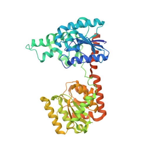
Sucrose phosphate synthase (SPS) catalyzes the transfer of a glycosyl group from an activated donor sugar, such as uridine diphosphate glucose (UDP-Glc), to a saccharide acceptor D-fructose 6-phosphate (F6P), resulting in the formation of UDP and D-sucrose-6'-phosphate (S6P). This is a central regulatory process in the production of sucrose in plants, cyanobacteria, and proteobacteria. Here, we report the crystal structure of SPS from the nonphotosynthetic bacterium Halothermothrix orenii and its complexes with the substrate F6P and the product S6P. SPS has two distinct Rossmann-fold domains with a large substrate binding cleft at the interdomain interface. Structures of two complexes show that both the substrate F6P and the product S6P bind to the A-domain of SPS. Based on comparative analysis of the SPS structure with other related enzymes, the donor substrate, nucleotide diphosphate glucose, binds to the B-domain of SPS. Furthermore, we propose a mechanism of catalysis by H. orenii SPS. Our findings indicate that SPS from H. orenii may represent a valid model for the catalytic domain of plant SPSs and thus may provide useful insight into the reaction mechanism of the plant enzyme.
Organizational Affiliation:
Department of Biological Sciences, National University of Singapore, Singapore 117543.