2R3J

Crystal Structure of Cyclin-Dependent Kinase 2 with inhibitor


Experimental Data Snapshot

  • Method: X-RAY DIFFRACTION
  • Resolution: 1.65 Å
  • R-Value Free: 0.236 
  • R-Value Work: 0.198 
  • R-Value Observed: 0.200 

wwPDB Validation   3D Report Full Report


Ligand Structure Quality Assessment 


This is version 1.1 of the entry. See complete history


Literature

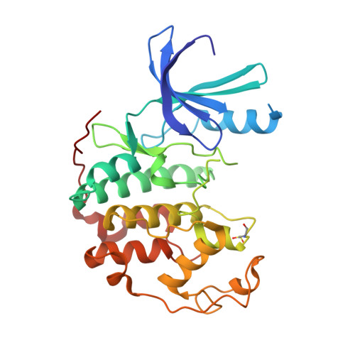
Structure-guided discovery of cyclin-dependent kinase inhibitors.

Fischmann, T.O.Hruza, A.Duca, J.S.Ramanathan, L.Mayhood, T.Windsor, W.T.Le, H.V.Guzi, T.J.Dwyer, M.P.Paruch, K.Doll, R.J.Lees, E.Parry, D.Seghezzi, W.Madison, V.

(2008) Biopolymers 89: 372-379

  • DOI: https://doi.org/10.1002/bip.20868
  • Primary Citation of Related Structures:  
    2R3F, 2R3G, 2R3H, 2R3I, 2R3J, 2R3K, 2R3L, 2R3M, 2R3N, 2R3O, 2R3P, 2R3Q, 2R3R

  • PubMed Abstract: 

    CDK2 inhibitors containing the related bicyclic heterocycles pyrazolopyrimidines and imidazopyrazines were discovered through high-throughput screening. Crystal structures of inhibitors with these bicyclic cores and two more related ones show that all but one have a common binding mode featuring two hydrogen bonds (H-bonds) to the backbone of the kinase hinge region. Even though ab initio computations indicated that the imidazopyrazine core would bind more tightly to the hinge, pyrazolopyrimidines gain an advantage in potency through participation of N4 in an H-bond network involving two catalytic residues and bridging water molecules. Further insight into inhibitor/CDK2 interactions was gained from analysis of additional crystal structures. Significant gains in potency were obtained by optimizing the fit of hydrophobic substituents to the gatekeeper region of the ATP binding site. The most potent inhibitors have good selectivity.


  • Organizational Affiliation

    Schering-Plough Research Institute, Kenilworth, NJ 07033, USA.


Macromolecules
Find similar proteins by:  (by identity cutoff)  |  3D Structure
Entity ID: 1
MoleculeChains Sequence LengthOrganismDetailsImage
Cell division protein kinase 2299Homo sapiensMutation(s): 0 
Gene Names: CDK2
EC: 2.7.11.22
UniProt & NIH Common Fund Data Resources
Find proteins for P24941 (Homo sapiens)
Explore P24941 
Go to UniProtKB:  P24941
PHAROS:  P24941
GTEx:  ENSG00000123374 
Entity Groups  
Sequence Clusters30% Identity50% Identity70% Identity90% Identity95% Identity100% Identity
UniProt GroupP24941
Sequence Annotations
Expand
  • Reference Sequence
Small Molecules
Ligands 1 Unique
IDChains Name / Formula / InChI Key2D Diagram3D Interactions
SCJ
Query on SCJ

Download Ideal Coordinates CCD File 
B [auth A]3-bromo-5-phenyl-N-(pyridin-3-ylmethyl)pyrazolo[1,5-a]pyrimidin-7-amine
C18 H14 Br N5
SMIZFGZXFDGISG-UHFFFAOYSA-N
Modified Residues  1 Unique
IDChains TypeFormula2D DiagramParent
CSD
Query on CSD
A
L-PEPTIDE LINKINGC3 H7 N O4 SCYS
Binding Affinity Annotations 
IDSourceBinding Affinity
SCJ Binding MOAD:  2R3J IC50: 10 (nM) from 1 assay(s)
PDBBind:  2R3J IC50: 10 (nM) from 1 assay(s)
Experimental Data & Validation

Experimental Data

  • Method: X-RAY DIFFRACTION
  • Resolution: 1.65 Å
  • R-Value Free: 0.236 
  • R-Value Work: 0.198 
  • R-Value Observed: 0.200 
  • Space Group: P 21 21 21
Unit Cell:
Length ( Å )Angle ( ˚ )
a = 53.5α = 90
b = 72.07β = 90
c = 72.1γ = 90
Software Package:
Software NamePurpose
DENZOdata reduction
SCALEPACKdata scaling
PDB_EXTRACTdata extraction
BUSTER-TNTrefinement

Structure Validation

View Full Validation Report


Ligand Structure Quality Assessment 


Entry History 

Deposition Data

Revision History  (Full details and data files)

  • Version 1.0: 2008-01-22
    Type: Initial release
  • Version 1.1: 2011-07-13
    Changes: Version format compliance