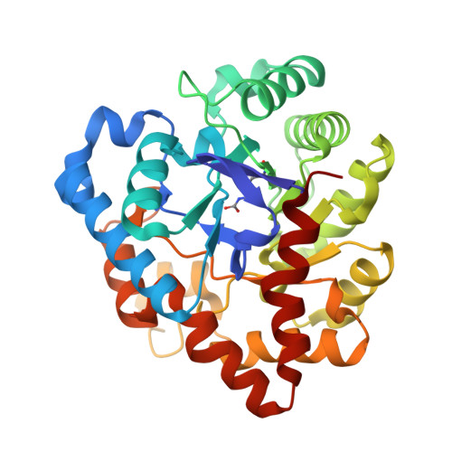
In crystallo capture of a Michaelis complex and product-binding modes of a bacterial phosphotriesterase
Jackson, C.J., Foo, J.L., Kim, H.K., Carr, P.D., Liu, J.W., Salem, G., Ollis, D.L.(2008) J Mol Biol 375: 1189-1196
- PubMed: 18082180
- DOI: https://doi.org/10.1016/j.jmb.2007.10.061
- Primary Citation of Related Structures:
2R1K, 2R1L, 2R1M, 2R1N, 2R1P, 3C86 - PubMed Abstract:
The mechanism by which the binuclear metallophosphotriesterases (PTEs, E.C. 3.1.8.1) catalyse substrate hydrolysis has been extensively studied. The mu-hydroxo bridge between the metal ions has been proposed to be the initiating nucleophile in the hydrolytic reaction. In contrast, analysis of some biomimetic systems has indicated that mu-hydroxo bridges are often not themselves nucleophiles, but act as general bases for freely exchangeable nucleophilic water molecules. Herein, we present crystallographic analyses of a bacterial PTE from Agrobacterium radiobacter, OpdA, capturing the enzyme-substrate complex during hydrolysis. This model of the Michaelis complex suggests the alignment of the substrate will favour attack from a solvent molecule terminally coordinated to the alpha-metal ion. The bridging of both metal ions by the product, without disruption of the mu-hydroxo bridge, is also consistent with nucleophilic attack occurring from the terminal position. When phosphodiesters are soaked into crystals of OpdA, they coordinate bidentately to the beta-metal ion, displacing the mu-hydroxo bridge. Thus, alternative product-binding modes exist for the PTEs, and it is the bridging mode that appears to result from phosphotriester hydrolysis. Kinetic analysis of the PTE and promiscuous phosphodiesterase activities confirms that the presence of a mu-hydroxo bridge during phosphotriester hydrolysis is correlated with a lower pK(a) for the nucleophile, consistent with a general base function during catalysis.
Organizational Affiliation:
Research School of Chemistry, Australian National University, Australian Capital Territory 0200, Australia.