2R15

Crystal structure of the myomesin domains 12 and 13


Experimental Data Snapshot

  • Method: X-RAY DIFFRACTION
  • Resolution: 2.24 Å
  • R-Value Free: 0.223 
  • R-Value Work: 0.196 

wwPDB Validation   3D Report Full Report


This is version 1.3 of the entry. See complete history


Literature

Molecular basis of the C-terminal tail-to-tail assembly of the sarcomeric filament protein myomesin.

Pinotsis, N.Lange, S.Perriard, J.C.Svergun, D.I.Wilmanns, M.

(2008) EMBO J 27: 253-264

  • DOI: https://doi.org/10.1038/sj.emboj.7601944
  • Primary Citation of Related Structures:  
    2R15

  • PubMed Abstract: 

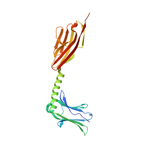
    Sarcomeric filament proteins display extraordinary properties in terms of protein length and mechanical elasticity, requiring specific anchoring and assembly mechanisms. To establish the molecular basis of terminal filament assembly, we have selected the sarcomeric M-band protein myomesin as a prototypic filament model. The crystal structure of the myomesin C-terminus, comprising a tandem array of two immunoglobulin (Ig) domains My12 and My13, reveals a dimeric end-to-end filament of 14.3 nm length. Although the two domains share the same fold, an unexpected rearrangement of one beta-strand reveals how they are evolved into unrelated functions, terminal filament assembly (My13) and filament propagation (My12). The two domains are connected by a six-turn alpha-helix, of which two turns are void of any interactions with other protein parts. Thus, the overall structure of the assembled myomesin C-terminus resembles a three-body beads-on-the-string model with potentially elastic properties. We predict that the found My12-helix-My13 domain topology may provide a structural template for the filament architecture of the entire C-terminal Ig domain array My9-My13 of myomesin.


  • Organizational Affiliation

    EMBL-Hamburg c/o DESY, Hamburg, Germany.


Macromolecules
Find similar proteins by:  (by identity cutoff)  |  3D Structure
Entity ID: 1
MoleculeChains Sequence LengthOrganismDetailsImage
Myomesin-1
A, B
212Homo sapiensMutation(s): 0 
Gene Names: MYOM1
UniProt & NIH Common Fund Data Resources
Find proteins for P52179 (Homo sapiens)
Explore P52179 
Go to UniProtKB:  P52179
PHAROS:  P52179
GTEx:  ENSG00000101605 
Entity Groups  
Sequence Clusters30% Identity50% Identity70% Identity90% Identity95% Identity100% Identity
UniProt GroupP52179
Sequence Annotations
Expand
  • Reference Sequence
Experimental Data & Validation

Experimental Data

  • Method: X-RAY DIFFRACTION
  • Resolution: 2.24 Å
  • R-Value Free: 0.223 
  • R-Value Work: 0.196 
  • Space Group: C 2 2 21
Unit Cell:
Length ( Å )Angle ( ˚ )
a = 61.504α = 90
b = 87.87β = 90
c = 203.081γ = 90
Software Package:
Software NamePurpose
CNSrefinement
REFMACrefinement
MAR345data collection
DENZOdata reduction
SCALEPACKdata scaling
CNSphasing

Structure Validation

View Full Validation Report


Entry History 

Deposition Data

Revision History  (Full details and data files)

  • Version 1.0: 2008-01-29
    Type: Initial release
  • Version 1.1: 2011-07-13
    Changes: Advisory, Version format compliance
  • Version 1.2: 2017-10-25
    Changes: Refinement description
  • Version 1.3: 2024-03-13
    Changes: Data collection, Database references, Derived calculations