2R0D

Crystal Structure of Autoinhibited Form of Grp1 Arf GTPase Exchange Factor


Experimental Data Snapshot

  • Method: X-RAY DIFFRACTION
  • Resolution: 2.04 Å
  • R-Value Free: 0.242 
  • R-Value Work: 0.208 
  • R-Value Observed: 0.210 

wwPDB Validation   3D Report Full Report


Ligand Structure Quality Assessment 


This is version 1.3 of the entry. See complete history


Literature

Structural Basis and Mechanism of Autoregulation in 3-Phosphoinositide-Dependent Grp1 Family Arf GTPase Exchange Factors.

DiNitto, J.P.Delprato, A.Gabe Lee, M.T.Cronin, T.C.Huang, S.Guilherme, A.Czech, M.P.Lambright, D.G.

(2007) Mol Cell 28: 569-583

  • DOI: https://doi.org/10.1016/j.molcel.2007.09.017
  • Primary Citation of Related Structures:  
    2R09, 2R0D

  • PubMed Abstract: 

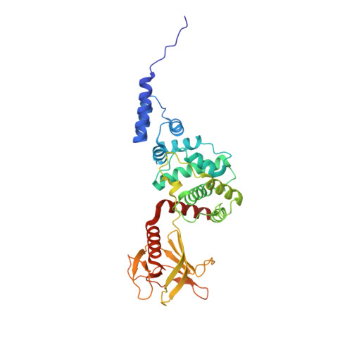
    Arf GTPases regulate membrane trafficking and actin dynamics. Grp1, ARNO, and Cytohesin-1 comprise a family of phosphoinositide-dependent Arf GTPase exchange factors with a Sec7-pleckstrin homology (PH) domain tandem. Here, we report that the exchange activity of the Sec7 domain is potently autoinhibited by conserved elements proximal to the PH domain. The crystal structure of the Grp1 Sec7-PH tandem reveals a pseudosubstrate mechanism of autoinhibition in which the linker region between domains and a C-terminal amphipathic helix physically block the docking sites for the switch regions of Arf GTPases. Mutations within either element result in partial or complete activation. Critical determinants of autoinhibition also contribute to insulin-stimulated plasma membrane recruitment. Autoinhibition can be largely reversed by binding of active Arf6 to Grp1 and by phosphorylation of tandem PKC sites in Cytohesin-1. These observations suggest that Grp1 family GEFs are autoregulated by mechanisms that depend on plasma membrane recruitment for activation.


  • Organizational Affiliation

    Program in Molecular Medicine, University of Massachusetts Medical School, Worcester, MA 01605, USA.


Macromolecules
Find similar proteins by:  (by identity cutoff)  |  3D Structure
Entity ID: 1
MoleculeChains Sequence LengthOrganismDetailsImage
Cytohesin-3
A, B
347Mus musculusMutation(s): 1 
Gene Names: Pscd3Grp1
UniProt
Find proteins for O08967 (Mus musculus)
Explore O08967 
Go to UniProtKB:  O08967
Entity Groups  
Sequence Clusters30% Identity50% Identity70% Identity90% Identity95% Identity100% Identity
UniProt GroupO08967
Sequence Annotations
Expand
  • Reference Sequence
Small Molecules
Ligands 3 Unique
IDChains Name / Formula / InChI Key2D Diagram3D Interactions
4IP
Query on 4IP

Download Ideal Coordinates CCD File 
F [auth A],
J [auth B]
INOSITOL-(1,3,4,5)-TETRAKISPHOSPHATE
C6 H16 O18 P4
CIPFCGZLFXVXBG-CNWJWELYSA-N
PEG
Query on PEG

Download Ideal Coordinates CCD File 
G [auth A],
H [auth A],
K [auth B]
DI(HYDROXYETHYL)ETHER
C4 H10 O3
MTHSVFCYNBDYFN-UHFFFAOYSA-N
SO4
Query on SO4

Download Ideal Coordinates CCD File 
C [auth A],
D [auth A],
E [auth A],
I [auth B]
SULFATE ION
O4 S
QAOWNCQODCNURD-UHFFFAOYSA-L
Binding Affinity Annotations 
IDSourceBinding Affinity
4IP BindingDB:  2R0D Kd: 100 (nM) from 1 assay(s)
Experimental Data & Validation

Experimental Data

  • Method: X-RAY DIFFRACTION
  • Resolution: 2.04 Å
  • R-Value Free: 0.242 
  • R-Value Work: 0.208 
  • R-Value Observed: 0.210 
  • Space Group: P 21 21 21
Unit Cell:
Length ( Å )Angle ( ˚ )
a = 82.906α = 90
b = 94.705β = 90
c = 115.436γ = 90
Software Package:
Software NamePurpose
REFMACrefinement
HKL-2000data collection
DENZOdata reduction
SCALEPACKdata scaling
PHASERphasing

Structure Validation

View Full Validation Report


Ligand Structure Quality Assessment 


Entry History 

Deposition Data

Revision History  (Full details and data files)

  • Version 1.0: 2007-12-04
    Type: Initial release
  • Version 1.1: 2011-07-13
    Changes: Derived calculations, Version format compliance
  • Version 1.2: 2021-10-20
    Changes: Database references, Derived calculations
  • Version 1.3: 2024-02-21
    Changes: Data collection