2QX0

Crystal Structure of Yersinia pestis HPPK (Ternary Complex)


Experimental Data Snapshot

  • Method: X-RAY DIFFRACTION
  • Resolution: 1.80 Å
  • R-Value Free: 0.269 
  • R-Value Work: 0.230 
  • R-Value Observed: 0.232 

wwPDB Validation   3D Report Full Report


Ligand Structure Quality Assessment 


This is version 1.3 of the entry. See complete history


Literature

Structure and activity of Yersinia pestis 6-hydroxymethyl-7,8-dihydropterin pyrophosphokinase as a novel target for the development of antiplague therapeutics.

Blaszczyk, J.Li, Y.Cherry, S.Alexandratos, J.Wu, Y.Shaw, G.Tropea, J.E.Waugh, D.S.Yan, H.Ji, X.

(2007) Acta Crystallogr D Biol Crystallogr 63: 1169-1177

  • DOI: https://doi.org/10.1107/S0907444907047452
  • Primary Citation of Related Structures:  
    2QX0

  • PubMed Abstract: 

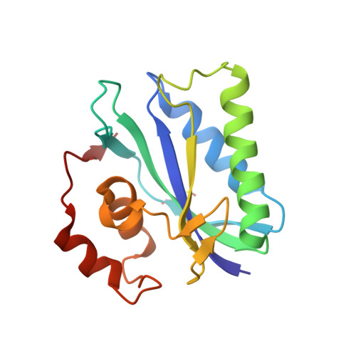
    6-Hydroxymethyl-7,8-dihydropterin pyrophosphokinase (HPPK) is a key enzyme in the folate-biosynthetic pathway and is essential for microorganisms but absent from mammals. HPPK catalyzes Mg(2+)-dependent pyrophosphoryl transfer from ATP to 6-hydroxymethyl-7,8-dihydropterin (HP). Previously, three-dimensional structures of Escherichia coli HPPK (EcHPPK) have been determined at almost every stage of its catalytic cycle and the reaction mechanism has been established. Here, the crystal structure of Yersinia pestis HPPK (YpHPPK) in complex with HP and an ATP analog is presented together with thermodynamic and kinetic characterizations. The two HPPK molecules differ significantly in a helix-loop area (alpha2-Lp3). YpHPPK has lower affinities than EcHPPK for both nucleotides and HP, but its rate constants for the mechanistic steps of both chemical transformation and product release are comparable with those of EcHPPK. Y. pestis, which causes plague, is a category A select agent according to the Centers for Disease Control and Prevention (CDC). Therefore, these structural and biochemical data are valuable for the design of novel medical countermeasures against plague.


  • Organizational Affiliation

    Macromolecular Crystallography Laboratory, National Cancer Institute, Frederick, MD 21702, USA.


Macromolecules
Find similar proteins by:  (by identity cutoff)  |  3D Structure
Entity ID: 1
MoleculeChains Sequence LengthOrganismDetailsImage
7,8-dihydro-6-hydroxymethylpterin-pyrophosphokinase
A, B
159Yersinia pestisMutation(s): 0 
Gene Names: folK
EC: 2.7.6.3
UniProt
Find proteins for A0A5P8YCA3 (Yersinia pestis)
Explore A0A5P8YCA3 
Go to UniProtKB:  A0A5P8YCA3
Entity Groups  
Sequence Clusters30% Identity50% Identity70% Identity90% Identity95% Identity100% Identity
UniProt GroupA0A5P8YCA3
Sequence Annotations
Expand
  • Reference Sequence
Small Molecules
Ligands 3 Unique
IDChains Name / Formula / InChI Key2D Diagram3D Interactions
APC
Query on APC

Download Ideal Coordinates CCD File 
E [auth A],
I [auth B]
DIPHOSPHOMETHYLPHOSPHONIC ACID ADENOSYL ESTER
C11 H18 N5 O12 P3
CAWZRIXWFRFUQB-IOSLPCCCSA-N
PH2
Query on PH2

Download Ideal Coordinates CCD File 
F [auth A],
J [auth B]
2-AMINO-6-HYDROXYMETHYL-7,8-DIHYDRO-3H-PTERIDIN-4-ONE
C7 H9 N5 O2
CQQNNQTXUGLUEV-UHFFFAOYSA-N
MG
Query on MG

Download Ideal Coordinates CCD File 
C [auth A],
D [auth A],
G [auth B],
H [auth B]
MAGNESIUM ION
Mg
JLVVSXFLKOJNIY-UHFFFAOYSA-N
Modified Residues  1 Unique
IDChains TypeFormula2D DiagramParent
CSO
Query on CSO
A, B
L-PEPTIDE LINKINGC3 H7 N O3 SCYS
Binding Affinity Annotations 
IDSourceBinding Affinity
PH2 PDBBind:  2QX0 Kd: 780 (nM) from 1 assay(s)
Binding MOAD:  2QX0 Kd: 780 (nM) from 1 assay(s)
Experimental Data & Validation

Experimental Data

  • Method: X-RAY DIFFRACTION
  • Resolution: 1.80 Å
  • R-Value Free: 0.269 
  • R-Value Work: 0.230 
  • R-Value Observed: 0.232 
  • Space Group: P 1 21 1
Unit Cell:
Length ( Å )Angle ( ˚ )
a = 43.4α = 90
b = 78.62β = 100.72
c = 52.6γ = 90
Software Package:
Software NamePurpose
AMoREphasing
CNSrefinement
PDB_EXTRACTdata extraction
ADSCdata collection
DENZOdata reduction
SCALEPACKdata scaling

Structure Validation

View Full Validation Report


Ligand Structure Quality Assessment 


Entry History 

Deposition Data

Revision History  (Full details and data files)

  • Version 1.0: 2007-10-23
    Type: Initial release
  • Version 1.1: 2011-07-13
    Changes: Version format compliance
  • Version 1.2: 2023-08-30
    Changes: Data collection, Database references, Derived calculations, Refinement description, Structure summary
  • Version 1.3: 2023-11-15
    Changes: Data collection