2QWO

Crystal structure of disulfide-bond-crosslinked complex of bovine hsc70 (1-394aa)R171C and bovine Auxilin (810-910aa)D876C in the ADP*Pi form #1


Experimental Data Snapshot

  • Method: X-RAY DIFFRACTION
  • Resolution: 1.70 Å
  • R-Value Free: 0.222 
  • R-Value Work: 0.193 
  • R-Value Observed: 0.194 

wwPDB Validation   3D Report Full Report


This is version 1.3 of the entry. See complete history


Literature

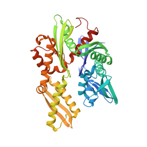
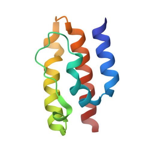
Structural basis of J cochaperone binding and regulation of Hsp70.

Jiang, J.Maes, E.G.Taylor, A.B.Wang, L.Hinck, A.P.Lafer, E.M.Sousa, R.

(2007) Mol Cell 28: 422-433

  • DOI: https://doi.org/10.1016/j.molcel.2007.08.022
  • Primary Citation of Related Structures:  
    2QW9, 2QWL, 2QWM, 2QWN, 2QWO, 2QWP, 2QWQ, 2QWR

  • PubMed Abstract: 

    The many protein processing reactions of the ATP-hydrolyzing Hsp70s are regulated by J cochaperones, which contain J domains that stimulate Hsp70 ATPase activity and accessory domains that present protein substrates to Hsp70s. We report the structure of a J domain complexed with a J responsive portion of a mammalian Hsp70. The J domain activates ATPase activity by directing the linker that connects the Hsp70 nucleotide binding domain (NBD) and substrate binding domain (SBD) toward a hydrophobic patch on the NBD surface. Binding of the J domain to Hsp70 displaces the SBD from the NBD, which may allow the SBD flexibility to capture diverse substrates. Unlike prokaryotic Hsp70, the SBD and NBD of the mammalian chaperone interact in the ADP state. Thus, although both nucleotides and J cochaperones modulate Hsp70 NBD:linker and NBD:SBD interactions, the intrinsic persistence of those interactions differs in different Hsp70s and this may optimize their activities for different cellular roles.


  • Organizational Affiliation

    Department of Biochemistry, University of Texas Health Science Center, San Antonio, TX 78229-3900, USA.


Macromolecules
Find similar proteins by:  (by identity cutoff)  |  3D Structure
Entity ID: 1
MoleculeChains Sequence LengthOrganismDetailsImage
Heat shock cognate 71 kDa protein394Bos taurusMutation(s): 1 
Gene Names: HSPA8HSC70
UniProt
Find proteins for P19120 (Bos taurus)
Explore P19120 
Go to UniProtKB:  P19120
Entity Groups  
Sequence Clusters30% Identity50% Identity70% Identity90% Identity95% Identity100% Identity
UniProt GroupP19120
Sequence Annotations
Expand
  • Reference Sequence
Find similar proteins by:  (by identity cutoff)  |  3D Structure
Entity ID: 2
MoleculeChains Sequence LengthOrganismDetailsImage
Putative tyrosine-protein phosphatase auxilin92Bos taurusMutation(s): 1 
Gene Names: DNAJC6
EC: 3.1.3.48
UniProt
Find proteins for Q27974 (Bos taurus)
Explore Q27974 
Go to UniProtKB:  Q27974
Entity Groups  
Sequence Clusters30% Identity50% Identity70% Identity90% Identity95% Identity100% Identity
UniProt GroupQ27974
Sequence Annotations
Expand
  • Reference Sequence
Small Molecules
Ligands 6 Unique
IDChains Name / Formula / InChI Key2D Diagram3D Interactions
ADP
Query on ADP

Download Ideal Coordinates CCD File 
G [auth A]ADENOSINE-5'-DIPHOSPHATE
C10 H15 N5 O10 P2
XTWYTFMLZFPYCI-KQYNXXCUSA-N
PO4
Query on PO4

Download Ideal Coordinates CCD File 
C [auth A]PHOSPHATE ION
O4 P
NBIIXXVUZAFLBC-UHFFFAOYSA-K
GOL
Query on GOL

Download Ideal Coordinates CCD File 
I [auth A],
J [auth A]
GLYCEROL
C3 H8 O3
PEDCQBHIVMGVHV-UHFFFAOYSA-N
ACY
Query on ACY

Download Ideal Coordinates CCD File 
H [auth A]ACETIC ACID
C2 H4 O2
QTBSBXVTEAMEQO-UHFFFAOYSA-N
MG
Query on MG

Download Ideal Coordinates CCD File 
F [auth A]MAGNESIUM ION
Mg
JLVVSXFLKOJNIY-UHFFFAOYSA-N
NA
Query on NA

Download Ideal Coordinates CCD File 
D [auth A],
E [auth A]
SODIUM ION
Na
FKNQFGJONOIPTF-UHFFFAOYSA-N
Binding Affinity Annotations 
IDSourceBinding Affinity
ADP BindingDB:  2QWO Kd: min: 320, max: 324 (nM) from 2 assay(s)
IC50: 280 (nM) from 1 assay(s)
Experimental Data & Validation

Experimental Data

  • Method: X-RAY DIFFRACTION
  • Resolution: 1.70 Å
  • R-Value Free: 0.222 
  • R-Value Work: 0.193 
  • R-Value Observed: 0.194 
  • Space Group: P 21 21 21
Unit Cell:
Length ( Å )Angle ( ˚ )
a = 38.698α = 90
b = 58.543β = 90
c = 226.236γ = 90
Software Package:
Software NamePurpose
d*TREKdata scaling
DENZOdata reduction
SCALEPACKdata scaling
MOLREPphasing
REFMACrefinement
PDB_EXTRACTdata extraction
d*TREKdata reduction

Structure Validation

View Full Validation Report


Entry History 

Deposition Data

Revision History  (Full details and data files)

  • Version 1.0: 2007-12-18
    Type: Initial release
  • Version 1.1: 2011-07-13
    Changes: Non-polymer description, Version format compliance
  • Version 1.2: 2017-10-25
    Changes: Refinement description
  • Version 1.3: 2021-10-20
    Changes: Database references, Derived calculations