2QV6

GTP cyclohydrolase III from M. jannaschii (MJ0145) complexed with GTP and metal ions


Experimental Data Snapshot

  • Method: X-RAY DIFFRACTION
  • Resolution: 2.00 Å
  • R-Value Free: 0.244 
  • R-Value Work: 0.206 
  • R-Value Observed: 0.208 

wwPDB Validation   3D Report Full Report


Ligand Structure Quality Assessment 


This is version 1.2 of the entry. See complete history


Literature

A New Use for a Familiar Fold: The X-ray Crystal Structure of GTP-Bound GTP Cyclohydrolase III from Methanocaldococcus jannaschii Reveals a Two Metal Ion Catalytic Mechanism

Morrison, S.D.Roberts, S.A.Zegeer, A.M.Montfort, W.R.Bandarian, V.

(2008) Biochemistry 47: 230-242

  • DOI: https://doi.org/10.1021/bi701782e
  • Primary Citation of Related Structures:  
    2QV6

  • PubMed Abstract: 

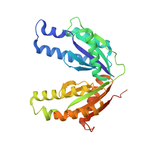
    GTP cyclohydrolase (GCH) III from Methanocaldococcus jannaschii, which catalyzes the conversion of GTP to 2-amino-5-formylamino-6-ribosylamino-4(3H)-pyrimidinone 5'-phosphate (FAPy), has been shown to require Mg2+ for catalytic activity and is activated by monovalent cations such as K+ and ammonium [Graham, D. E., Xu, H., and White, R. H. (2002) Biochemistry 41, 15074-15084]. The reaction is formally identical to that catalyzed by a GCH II ortholog (SCO 6655) from Streptomyces coelicolor; however, SCO 6655, like other GCH II proteins, is a zinc-containing protein. The structure of GCH III complexed with GTP solved at 2 A resolution clearly shows that GCH III adopts a distinct fold that is closely related to the palm domains of phosphodiesterases, such as DNA polymerase I. GCH III is a tetramer of identical subunits; each monomer is composed of an N- and a C-terminal domain that adopt nearly superimposible structures, suggesting that the protein has arisen by gene duplication. Three metal ions were located in the active site, two of which occupy positions that are analogous to those occupied by divalent metal ions in the structures of a number of palm domain containing proteins, such as DNA polymerase I. Two conserved Asp residues that coordinate the metal ions, which are also found in palm domain containing proteins, are observed in GCH III. Site-directed variants (Asp-->Asn) of these residues in GCH III are less active than wild-type. The third metal ion, most likely a potassium ion, is involved in substrate recognition through coordination of O6 of GTP. The arrangement of the metal ions in the active site suggests that GCH III utilizes two metal ion catalysis. The structure of GCH III extends the repertoire of possible reactions with a palm fold to include cyclohydrolase chemistry.


  • Organizational Affiliation

    Department of Biochemistry and Molecular Biophysics, The University of Arizona, Tucson, Arizona 85721, USA.


Macromolecules
Find similar proteins by:  (by identity cutoff)  |  3D Structure
Entity ID: 1
MoleculeChains Sequence LengthOrganismDetailsImage
GTP cyclohydrolase III
A, B, C, D
268Methanocaldococcus jannaschiiMutation(s): 0 
Gene Names: gch3
EC: 3.5.4.29
UniProt
Find proteins for Q57609 (Methanocaldococcus jannaschii (strain ATCC 43067 / DSM 2661 / JAL-1 / JCM 10045 / NBRC 100440))
Explore Q57609 
Go to UniProtKB:  Q57609
Entity Groups  
Sequence Clusters30% Identity50% Identity70% Identity90% Identity95% Identity100% Identity
UniProt GroupQ57609
Sequence Annotations
Expand
  • Reference Sequence
Small Molecules
Ligands 4 Unique
IDChains Name / Formula / InChI Key2D Diagram3D Interactions
GTP
Query on GTP

Download Ideal Coordinates CCD File 
H [auth A],
L [auth B],
P [auth C],
T [auth D]
GUANOSINE-5'-TRIPHOSPHATE
C10 H16 N5 O14 P3
XKMLYUALXHKNFT-UUOKFMHZSA-N
CA
Query on CA

Download Ideal Coordinates CCD File 
F [auth A],
J [auth B],
N [auth C],
R [auth D]
CALCIUM ION
Ca
BHPQYMZQTOCNFJ-UHFFFAOYSA-N
K
Query on K

Download Ideal Coordinates CCD File 
E [auth A],
I [auth B],
M [auth C],
Q [auth D]
POTASSIUM ION
K
NPYPAHLBTDXSSS-UHFFFAOYSA-N
NA
Query on NA

Download Ideal Coordinates CCD File 
G [auth A],
K [auth B],
O [auth C],
S [auth D]
SODIUM ION
Na
FKNQFGJONOIPTF-UHFFFAOYSA-N
Experimental Data & Validation

Experimental Data

  • Method: X-RAY DIFFRACTION
  • Resolution: 2.00 Å
  • R-Value Free: 0.244 
  • R-Value Work: 0.206 
  • R-Value Observed: 0.208 
  • Space Group: P 21 21 2
Unit Cell:
Length ( Å )Angle ( ˚ )
a = 100.203α = 90
b = 129.578β = 90
c = 90.876γ = 90
Software Package:
Software NamePurpose
CrystalCleardata collection
SOLVEphasing
REFMACrefinement
CrystalCleardata reduction
CrystalCleardata scaling

Structure Validation

View Full Validation Report


Ligand Structure Quality Assessment 


Entry History 

Deposition Data

Revision History  (Full details and data files)

  • Version 1.0: 2008-01-15
    Type: Initial release
  • Version 1.1: 2011-07-13
    Changes: Version format compliance
  • Version 1.2: 2024-02-21
    Changes: Data collection, Database references, Derived calculations