Molecular determinants of substrate specificity in plant 5'-methylthioadenosine nucleosidases.
Siu, K.K., Lee, J.E., Sufrin, J.R., Moffatt, B.A., McMillan, M., Cornell, K.A., Isom, C., Howell, P.L.(2008) J Mol Biol 378: 112-128
- PubMed: 18342331
- DOI: https://doi.org/10.1016/j.jmb.2008.01.088
- Primary Citation of Related Structures:
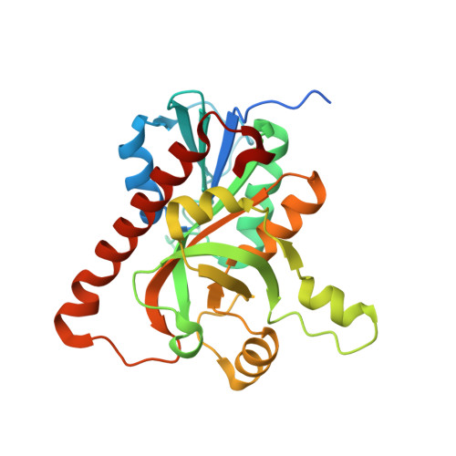
2QSU, 2QTG, 2QTT - PubMed Abstract:
5'-Methylthioadenosine (MTA)/S-adenosylhomocysteine (SAH) nucleosidase (MTAN) is essential for cellular metabolism and development in many bacterial species. While the enzyme is found in plants, plant MTANs appear to select for MTA preferentially, with little or no affinity for SAH. To understand what determines substrate specificity in this enzyme, MTAN homologues from Arabidopsis thaliana (AtMTAN1 and AtMTAN2, which are referred to as AtMTN1 and AtMTN2 in the plant literature) have been characterized kinetically. While both homologues hydrolyze MTA with comparable kinetic parameters, only AtMTAN2 shows activity towards SAH. AtMTAN2 also has higher catalytic activity towards other substrate analogues with longer 5'-substituents. The structures of apo AtMTAN1 and its complexes with the substrate- and transition-state-analogues, 5'-methylthiotubercidin and formycin A, respectively, have been determined at 2.0-1.8 A resolution. A homology model of AtMTAN2 was generated using the AtMTAN1 structures. Comparison of the AtMTAN1 and AtMTAN2 structures reveals that only three residues in the active site differ between the two enzymes. Our analysis suggests that two of these residues, Leu181/Met168 and Phe148/Leu135 in AtMTAN1/AtMTAN2, likely account for the divergence in specificity of the enzymes. Comparison of the AtMTAN1 and available Escherichia coli MTAN (EcMTAN) structures suggests that a combination of differences in the 5'-alkylthio binding region and reduced conformational flexibility in the AtMTAN1 active site likely contribute to its reduced efficiency in binding substrate analogues with longer 5'-substituents. In addition, in contrast to EcMTAN, the active site of AtMTAN1 remains solvated in its ligand-bound forms. As the apparent pK(a) of an amino acid depends on its local environment, the putative catalytic acid Asp225 in AtMTAN1 may not be protonated at physiological pH and this suggests the transition state of AtMTAN1, like human MTA phosphorylase and Streptococcus pneumoniae MTAN, may be different from that found in EcMTAN.
Organizational Affiliation:
Program in Molecular Structure and Function, Research Institute, Hospital for Sick Children, 555 University Avenue, Toronto, Ontario, Canada.