Effector Proteins Exert an Important Influence on the Signaling-active State of the Small GTPase Cdc42.
Phillips, M.J., Calero, G., Chan, B., Ramachandran, S., Cerione, R.A.(2008) J Biol Chem 283: 14153-14164
- PubMed: 18348980
- DOI: https://doi.org/10.1074/jbc.M706271200
- Primary Citation of Related Structures:
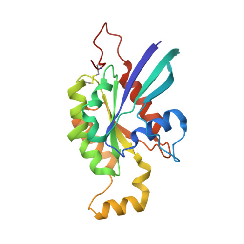
2QRZ - PubMed Abstract:
GTP-binding (G) proteins regulate the flow of information in cellular signaling pathways by alternating between a GTP-bound "active" state and a GDP-bound "inactive" state. Cdc42, a member of the Rho family of Ras-related small G-proteins, plays key roles in the regulation of cell shape, motility, and growth. Here we describe the high resolution x-ray crystal structure for Cdc42 bound to the GTP analog guanylyl beta,gamma-methylene-diphosphonate (GMP-PCP) (i.e. the presumed signaling-active state) and show that it is virtually identical to the structures for the signaling-inactive, GDP-bound form of the protein, contrary to what has been reported for Ras and other G-proteins. Especially surprising was that the GMP-PCP- and GDP-bound forms of Cdc42 did not show detectable differences in their Switch I and Switch II loops. Fluorescence studies using a Cdc42 mutant in which a tryptophan residue was introduced at position 32 of Switch I also showed that there was little difference in the Switch I conformation between the GDP- and GMP-PCP-bound states (i.e. <10%), which again differed from Ras where much larger changes in Trp-32 fluorescence were observed when comparing these two nucleotide-bound states (>30%). However, the binding of an effector protein induced significant changes in the Trp-32 emission specifically from GMP-PCP-bound Cdc42, as well as in the phosphate resonances for GTP bound to this G-protein as indicated in NMR studies. An examination of the available structures for Cdc42 complexed to different effector proteins, versus the x-ray crystal structure for GMP-PCP-bound Cdc42, provides a possible explanation for how effectors can distinguish between the GTP- and GDP-bound forms of this G-protein and ensure that the necessary conformational changes for signal propagation occur.
Organizational Affiliation:
Department of Chemistry and Chemical Biology, Baker Laboratory, Cornell University, Ithaca, New York 14853, USA.