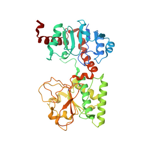
2QRL

Crystal Structure of Oxalylglycine-bound Saccharopine Dehydrogenase (L-Lys Forming) from Saccharomyces cerevisiae


Experimental Data Snapshot

  • Method: X-RAY DIFFRACTION
  • Resolution: 1.60 Å
  • R-Value Free: 0.241 
  • R-Value Work: 0.208 
  • R-Value Observed: 0.209 

wwPDB Validation   3D Report Full Report


This is version 1.3 of the entry. See complete history


Literature

Crystal Structures of Ligand-Bound Saccharopine Dehydrogenase from Saccharomyces cerevisiae

Andi, B.Xu, H.Cook, P.F.West, A.H.

(2007) Biochemistry 46: 12512-12521

  • DOI: https://doi.org/10.1021/bi701428m
  • Primary Citation of Related Structures:  
    2QRJ, 2QRK, 2QRL

  • PubMed Abstract: 

    Three structures of saccharopine dehydrogenase (l-lysine-forming) (SDH) have been determined in the presence of sulfate, adenosine monophosphate (AMP), and oxalylglycine (OxGly). In the sulfate-bound structure, a sulfate ion binds in a cleft between the two domains of SDH, occupies one of the substrate carboxylate binding sites, and results in partial closure of the active site of the enzyme due to a domain rotation of almost 12 degrees in comparison to the apoenzyme structure. In the second structure, AMP binds to the active site in an area where the NAD+ cofactor is expected to bind. All of the AMP moieties (adenine ring, ribose, and phosphate) interact with specific residues of the enzyme. In the OxGly-bound structure, carboxylates of OxGly interact with arginine residues representative of the manner in which substrate (alpha-ketoglutarate and saccharopine) may bind. The alpha-keto group of OxGly interacts with Lys77 and His96, which are candidates for acid-base catalysis. Analysis of ligand-enzyme interactions, comparative structural analysis, corroboration with kinetic data, and discussion of a ternary complex model are presented in this study.


  • Organizational Affiliation

    Department of Chemistry and Biochemistry, University of Oklahoma, 620 Parrington Oval, Norman, Oklahoma 73019, USA.


Macromolecules
Find similar proteins by:  (by identity cutoff)  |  3D Structure
Entity ID: 1
MoleculeChains Sequence LengthOrganismDetailsImage
Saccharopine dehydrogenase, NAD+, L-lysine-forming394Saccharomyces cerevisiaeMutation(s): 0 
Gene Names: LYS1
EC: 1.5.1.7
UniProt
Find proteins for P38998 (Saccharomyces cerevisiae (strain ATCC 204508 / S288c))
Explore P38998 
Go to UniProtKB:  P38998
Entity Groups  
Sequence Clusters30% Identity50% Identity70% Identity90% Identity95% Identity100% Identity
UniProt GroupP38998
Sequence Annotations
Expand
  • Reference Sequence
Small Molecules
Ligands 1 Unique
IDChains Name / Formula / InChI Key2D Diagram3D Interactions
OGA
Query on OGA

Download Ideal Coordinates CCD File 
B [auth A]N-OXALYLGLYCINE
C4 H5 N O5
BIMZLRFONYSTPT-UHFFFAOYSA-N
Binding Affinity Annotations 
IDSourceBinding Affinity
OGA PDBBind:  2QRL Ki: 1.00e+5 (nM) from 1 assay(s)
Binding MOAD:  2QRL Ki: 1.00e+5 (nM) from 1 assay(s)
Experimental Data & Validation

Experimental Data

  • Method: X-RAY DIFFRACTION
  • Resolution: 1.60 Å
  • R-Value Free: 0.241 
  • R-Value Work: 0.208 
  • R-Value Observed: 0.209 
  • Space Group: P 21 21 21
Unit Cell:
Length ( Å )Angle ( ˚ )
a = 64.762α = 90
b = 74.301β = 90
c = 74.619γ = 90
Software Package:
Software NamePurpose
d*TREKdata scaling
PHASERphasing
REFMACrefinement
PDB_EXTRACTdata extraction
CrystalCleardata collection
d*TREKdata reduction
ARP/wARPmodel building

Structure Validation

View Full Validation Report


Entry History 

Deposition Data

Revision History  (Full details and data files)

  • Version 1.0: 2007-10-30
    Type: Initial release
  • Version 1.1: 2011-07-13
    Changes: Version format compliance
  • Version 1.2: 2017-10-25
    Changes: Refinement description
  • Version 1.3: 2023-08-30
    Changes: Data collection, Database references, Derived calculations, Refinement description