


Crystal Structure of Ferritin like, Diiron-carboxylate Proteins from Bacillus anthracis str. Ames.
Kim, Y., Joachimiak, G., Wu, R., Patterson, S., Gornicki, P., Joachimiak, A.To be published.
Experimental Data Snapshot
wwPDB Validation 3D Report Full Report

Entity ID: 1 | |||||
|---|---|---|---|---|---|
| Molecule | Chains | Sequence Length | Organism | Details | Image |
| Sigma B operon | 149 | Bacillus anthracis str. Ames | Mutation(s): 0 Gene Names: BAS0929, BA_0993, GBAA0993 | 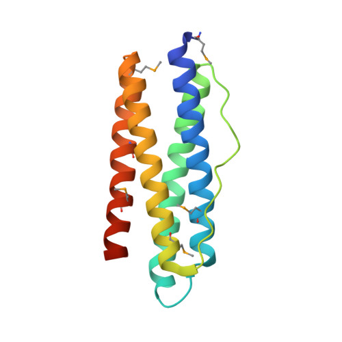 | |
UniProt | |||||
Find proteins for Q9K5J5 (Bacillus anthracis) Explore Q9K5J5 Go to UniProtKB: Q9K5J5 | |||||
Entity Groups | |||||
| Sequence Clusters | 30% Identity50% Identity70% Identity90% Identity95% Identity100% Identity | ||||
| UniProt Group | Q9K5J5 | ||||
Sequence AnnotationsExpand | |||||
| |||||
| Modified Residues 1 Unique | |||||
|---|---|---|---|---|---|
| ID | Chains | Type | Formula | 2D Diagram | Parent |
| MSE Query on MSE | A | L-PEPTIDE LINKING | C5 H11 N O2 Se |  | MET |
| Length ( Å ) | Angle ( ˚ ) |
|---|---|
| a = 100.225 | α = 90 |
| b = 100.225 | β = 90 |
| c = 100.225 | γ = 90 |
| Software Name | Purpose |
|---|---|
| REFMAC | refinement |
| HKL-3000 | data reduction |
| HKL-3000 | data scaling |
| HKL-3000 | phasing |
| SHELXCD | phasing |
| SHELXE | model building |
| MLPHARE | phasing |
| RESOLVE | phasing |
RCSB PDB (citation) is hosted by
RCSB PDB is a member of the

