Structure-based mechanism of ligand binding for periplasmic solute-binding protein of the Bug family.
Herrou, J., Bompard, C., Antoine, R., Leroy, A., Rucktooa, P., Hot, D., Huvent, I., Locht, C., Villeret, V., Jacob-Dubuisson, F.(2007) J Mol Biol 373: 954-964
- PubMed: 17870093
- DOI: https://doi.org/10.1016/j.jmb.2007.08.006
- Primary Citation of Related Structures:
2QPQ - PubMed Abstract:
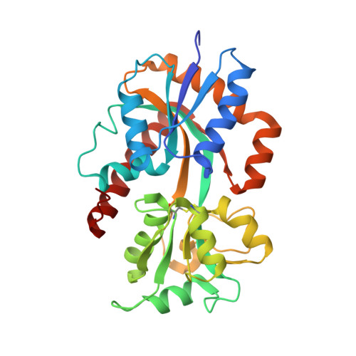
Bug proteins form a large family of periplasmic solute-binding proteins well represented in beta-proteobacteria. They adopt a characteristic Venus flytrap fold with two globular domains bisected by a ligand-binding cleft. The structures of two liganded Bug proteins have revealed that the family is specific for carboxylated solutes, with a characteristic mode of binding involving two highly conserved beta strand-beta turn-alpha helix motifs originating from each domain. These two motifs form hydrogen bonds with a carboxylate group of the ligand, both directly and via conserved water molecules, and have thus been termed the carboxylate pincers. In both crystallized Bug proteins, the ligands were found enclosed between the two domains and inaccessible to solvent, suggesting an inter-domain hinge-bending motion upon ligand binding. We report here the first structures of an open, unliganded Bug protein and of the same protein with a citrate ion bound in the open cavity. One of the ligand carboxylate groups is bound to one half of the carboxylate pincers by the beta strand-beta turn-alpha helix motif from domain 1, and the citrate ion forms several additional interactions with domain 1. The ligand is accessible to solvent and has very few contacts with domain 2. In this open, liganded structure, the second part of the carboxylate pincers originating from domain 2 is not stabilized by ligand binding, and a loop replaces the beta turn. In the unliganded structure, both motifs of the carboxylate pincers are highly mobile, and neither of the two beta turns is formed. Thus, ligand recognition is performed by domain 1, with the carboxylate group serving as an initial anchoring point. Stabilization of the closed conformation requires proper interactions to be established with domain 2, and thus domain 2 discriminates between productively and non-productively bound ligands.
Organizational Affiliation:
INSERM U629, 1 Rue du Pr Calmette, 59019 Lille cedex, France.