Structural Analysis of the Bacterial HPr Kinase/Phosphorylase V267F Mutant Gives Insights into the Allosteric Regulation Mechanism of This Bifunctional Enzyme.
Chaptal, V., Vincent, F., Gueguen-Chaignon, V., Monedero, V., Poncet, S., Deutscher, J., Nessler, S., Morera, S.(2007) J Biol Chem 282: 34952-34957
- PubMed: 17878158
- DOI: https://doi.org/10.1074/jbc.M705979200
- Primary Citation of Related Structures:
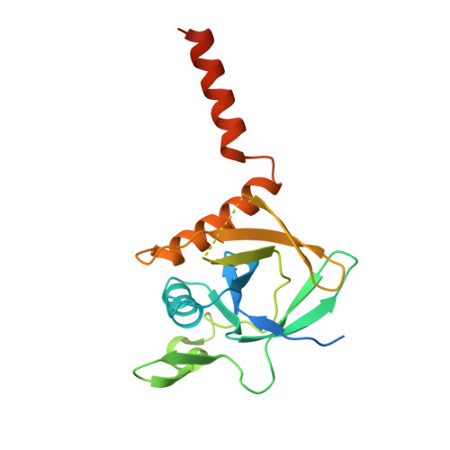
2QMH - PubMed Abstract:
The HPr kinase/phosphorylase (HPrK/P) is a bifunctional enzyme that controls the phosphorylation state of the phospho-carrier protein HPr, which regulates the utilization of carbon sources in Gram-positive bacteria. It uses ATP or pyrophosphate for the phosphorylation of serine 46 of HPr and inorganic phosphate for the dephosphorylation of Ser(P)-46-HPr via a phosphorolysis reaction. HPrK/P is a hexameric protein kinase of a new type with a catalytic core belonging to the family of nucleotide-binding protein with Walker A motif. It exhibits no structural similarity to eukaryotic protein kinases. So far, HPrK/P structures have shown the enzyme in its phosphorylase conformation. They permitted a detailed characterization of the phosphorolysis mechanism. In the absence of a structure with bound nucleotide, we used the V267F mutant enzyme to assess the kinase conformation. Indeed, the V267F replacement was found to cause an almost entire loss of the phosphorylase activity of Lactobacillus casei HPrK/P. In contrast, the kinase activity remained conserved. To elucidate the structural alterations leading to this drastic change of activity, the x-ray structure of the catalytic domain of L. casei HPrK/P-V267F was determined at 2.6A resolution. A comparison with the structure of the wild type enzyme showed that the mutation induces conformation changes compatible with the switch from phosphorylase to kinase function. Together with nucleotide binding fluorescence measurements, these results allowed us to decipher the cooperative behavior of the protein and to gain new insights into the allosteric regulation mechanism of HPrK/P.
Organizational Affiliation:
Laboratoire d'Enzymologie et Biochimie Structurales, CNRS, 1 Avenue de Terrasse, 91198 Gif-sur Yvette, France.