2QFK

X-ray Crystal Structure Analysis of the Binding Site in the Ferric and Oxyferrous Forms of the Recombinant Heme Dehaloperoxidase Cloned from Amphitrite ornata


Experimental Data Snapshot

  • Method: X-RAY DIFFRACTION
  • Resolution: 1.62 Å
  • R-Value Free: 0.239 
  • R-Value Work: 0.191 
  • R-Value Observed: 0.194 

wwPDB Validation   3D Report Full Report


Ligand Structure Quality Assessment 


This is version 1.3 of the entry. See complete history


Literature

X-ray crystal structural analysis of the binding site in the ferric and oxyferrous forms of the recombinant heme dehaloperoxidase cloned from Amphitrite ornata

de Serrano, V.S.Chen, Z.Davis, M.F.Franzen, S.

(2007) Acta Crystallogr D Biol Crystallogr 63: 1094-1101

  • DOI: https://doi.org/10.1107/S0907444907043417
  • Primary Citation of Related Structures:  
    2QFK, 2QFN

  • PubMed Abstract: 

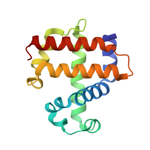
    The dehaloperoxidase (DHP) from the terebellid polychaete Amphitrite ornata is an enzyme that converts para-halogenated phenols to the corresponding quinones in the presence of hydrogen peroxide. Its enzymatic activity is similar to that of heme peroxidases such as horseradish peroxidase, yet it has the structural characteristics of the globin family of proteins, the main functions of which are oxygen transport and storage. In order to investigate the dual function of this hemoglobin peroxidase, the enzyme was expressed in Escherichia coli as a recombinant protein in its wild-type form and as a mutant protein in which Cys73 was replaced by a serine residue (C73S). Both the wild-type and mutant proteins were crystallized and their structures were determined at 100 K to a resolution of 1.62 A. The structure of the wild-type protein demonstrated that it was in the metaquo form, with the heme iron in the ferric oxidation state and the bound water lying 2.2 A from the heme iron. The structure of the C73S mutant protein was shown to contain a ferrous heme iron with a bound oxygen molecule. The bent bonding geometry of the Fe-O(1)-O(2) adduct results in a hydrogen bond of length 2.8 A between the second O atom, O(2), of molecular oxygen and N(2) of the distal histidine residue (His55) in both subunits contained within the asymmetric unit. This hydrogen-bonding interaction between His55 and the bound diatomic oxygen molecule provides new insight into the catalytic activation of H(2)O(2), which is essential for peroxidase activity.


  • Organizational Affiliation

    Department of Chemistry, North Carolina State University, Raleigh, NC, USA. vsserran@ncsu.edu


Macromolecules
Find similar proteins by:  (by identity cutoff)  |  3D Structure
Entity ID: 1
MoleculeChains Sequence LengthOrganismDetailsImage
Dehaloperoxidase A
A, B
137Amphitrite ornataMutation(s): 0 
Gene Names: dhpA
UniProt
Find proteins for Q9NAV8 (Amphitrite ornata)
Explore Q9NAV8 
Go to UniProtKB:  Q9NAV8
Entity Groups  
Sequence Clusters30% Identity50% Identity70% Identity90% Identity95% Identity100% Identity
UniProt GroupQ9NAV8
Sequence Annotations
Expand
  • Reference Sequence
Small Molecules
Ligands 3 Unique
IDChains Name / Formula / InChI Key2D Diagram3D Interactions
HEM
Query on HEM

Download Ideal Coordinates CCD File 
F [auth A],
J [auth B]
PROTOPORPHYRIN IX CONTAINING FE
C34 H32 Fe N4 O4
KABFMIBPWCXCRK-RGGAHWMASA-L
SO4
Query on SO4

Download Ideal Coordinates CCD File 
E [auth A],
H [auth B],
I [auth B]
SULFATE ION
O4 S
QAOWNCQODCNURD-UHFFFAOYSA-L
NH4
Query on NH4

Download Ideal Coordinates CCD File 
C [auth A],
D [auth A],
G [auth B]
AMMONIUM ION
H4 N
QGZKDVFQNNGYKY-UHFFFAOYSA-O
Experimental Data & Validation

Experimental Data

  • Method: X-RAY DIFFRACTION
  • Resolution: 1.62 Å
  • R-Value Free: 0.239 
  • R-Value Work: 0.191 
  • R-Value Observed: 0.194 
  • Space Group: P 21 21 21
Unit Cell:
Length ( Å )Angle ( ˚ )
a = 60.686α = 90
b = 67.572β = 90
c = 67.622γ = 90
Software Package:
Software NamePurpose
DENZOdata reduction
SCALEPACKdata scaling
PHASERphasing
REFMACrefinement
PDB_EXTRACTdata extraction
HKL-2000data collection
HKL-2000data reduction
HKL-2000data scaling

Structure Validation

View Full Validation Report


Ligand Structure Quality Assessment 


Entry History 

Deposition Data

Revision History  (Full details and data files)

  • Version 1.0: 2008-07-08
    Type: Initial release
  • Version 1.1: 2011-07-13
    Changes: Version format compliance
  • Version 1.2: 2017-10-18
    Changes: Refinement description
  • Version 1.3: 2024-02-21
    Changes: Data collection, Database references, Derived calculations