2QF7

Crystal structure of a complete multifunctional pyruvate carboxylase from Rhizobium etli


Experimental Data Snapshot

  • Method: X-RAY DIFFRACTION
  • Resolution: 2.00 Å
  • R-Value Free: 0.223 
  • R-Value Work: 0.178 
  • R-Value Observed: 0.180 

wwPDB Validation   3D Report Full Report


Ligand Structure Quality Assessment 


This is version 1.2 of the entry. See complete history


Literature

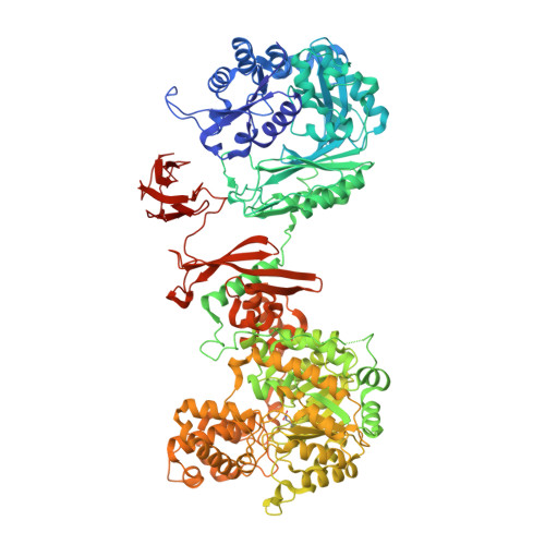
Domain architecture of pyruvate carboxylase, a biotin-dependent multifunctional enzyme

St Maurice, M.Reinhardt, L.Surinya, K.H.Attwood, P.V.Wallace, J.C.Cleland, W.W.Rayment, I.

(2007) Science 317: 1076-1079

  • DOI: https://doi.org/10.1126/science.1144504
  • Primary Citation of Related Structures:  
    2QF7

  • PubMed Abstract: 

    Biotin-dependent multifunctional enzymes carry out metabolically important carboxyl group transfer reactions and are potential targets for the treatment of obesity and type 2 diabetes. These enzymes use a tethered biotin cofactor to carry an activated carboxyl group between distantly spaced active sites. The mechanism of this transfer has remained poorly understood. Here we report the complete structure of pyruvate carboxylase at 2.0 angstroms resolution, which shows its domain arrangement. The structure, when combined with mutagenic analysis, shows that intermediate transfer occurs between active sites on separate polypeptide chains. In addition, domain rearrangements associated with activator binding decrease the distance between active-site pairs, providing a mechanism for allosteric activation. This description provides insight into the function of biotin-dependent enzymes and presents a new paradigm for multifunctional enzyme catalysis.


  • Organizational Affiliation

    Department of Biochemistry, University of Wisconsin, Madison, WI 53706, USA.


Macromolecules
Find similar proteins by:  (by identity cutoff)  |  3D Structure
Entity ID: 1
MoleculeChains Sequence LengthOrganismDetailsImage
Pyruvate carboxylase protein
A, B
1,165Rhizobium etli CFN 42Mutation(s): 1 
Gene Names: pyc
EC: 6.4.1.1
UniProt
Find proteins for Q2K340 (Rhizobium etli (strain CFN 42 / ATCC 51251))
Explore Q2K340 
Go to UniProtKB:  Q2K340
Entity Groups  
Sequence Clusters30% Identity50% Identity70% Identity90% Identity95% Identity100% Identity
UniProt GroupQ2K340
Sequence Annotations
Expand
  • Reference Sequence
Small Molecules
Ligands 7 Unique
IDChains Name / Formula / InChI Key2D Diagram3D Interactions
COA
Query on COA

Download Ideal Coordinates CCD File 
I [auth A]COENZYME A
C21 H36 N7 O16 P3 S
RGJOEKWQDUBAIZ-IBOSZNHHSA-N
AGS
Query on AGS

Download Ideal Coordinates CCD File 
J [auth A],
R [auth B]
PHOSPHOTHIOPHOSPHORIC ACID-ADENYLATE ESTER
C10 H16 N5 O12 P3 S
NLTUCYMLOPLUHL-KQYNXXCUSA-N
GOL
Query on GOL

Download Ideal Coordinates CCD File 
S [auth B]GLYCEROL
C3 H8 O3
PEDCQBHIVMGVHV-UHFFFAOYSA-N
ZN
Query on ZN

Download Ideal Coordinates CCD File 
E [auth A],
L [auth B]
ZINC ION
Zn
PTFCDOFLOPIGGS-UHFFFAOYSA-N
FMT
Query on FMT

Download Ideal Coordinates CCD File 
K [auth A],
T [auth B]
FORMIC ACID
C H2 O2
BDAGIHXWWSANSR-UHFFFAOYSA-N
CL
Query on CL

Download Ideal Coordinates CCD File 
G [auth A],
H [auth A],
P [auth B],
Q [auth B]
CHLORIDE ION
Cl
VEXZGXHMUGYJMC-UHFFFAOYSA-M
MG
Query on MG

Download Ideal Coordinates CCD File 
C [auth A]
D [auth A]
F [auth A]
M [auth B]
N [auth B]
C [auth A],
D [auth A],
F [auth A],
M [auth B],
N [auth B],
O [auth B]
MAGNESIUM ION
Mg
JLVVSXFLKOJNIY-UHFFFAOYSA-N
Modified Residues  1 Unique
IDChains TypeFormula2D DiagramParent
KCX
Query on KCX
A, B
L-PEPTIDE LINKINGC7 H14 N2 O4LYS
Experimental Data & Validation

Experimental Data

  • Method: X-RAY DIFFRACTION
  • Resolution: 2.00 Å
  • R-Value Free: 0.223 
  • R-Value Work: 0.178 
  • R-Value Observed: 0.180 
  • Space Group: C 1 2 1
Unit Cell:
Length ( Å )Angle ( ˚ )
a = 234.729α = 90
b = 93.258β = 107.33
c = 137.222γ = 90
Software Package:
Software NamePurpose
SOLVEphasing
RESOLVEphasing
REFMACrefinement
PDB_EXTRACTdata extraction
HKL-2000data collection
DENZOdata reduction
SCALEPACKdata scaling

Structure Validation

View Full Validation Report


Ligand Structure Quality Assessment 


Entry History 

Deposition Data

Revision History  (Full details and data files)

  • Version 1.0: 2007-09-04
    Type: Initial release
  • Version 1.1: 2011-07-13
    Changes: Advisory, Derived calculations, Source and taxonomy, Version format compliance
  • Version 1.2: 2012-02-15
    Changes: Non-polymer description