Biochemical and Structural Characterization of Pseudomonas aeruginosa Bfd and FPR: Ferredoxin NADP(+) Reductase and Not Ferredoxin Is the Redox Partner of Heme Oxygenase under Iron-Starvation Conditions
Wang, A., Zeng, Y., Han, H., Weeratunga, S., Morgan, B.N., Moenne-Loccoz, P., Schonbrunn, E., Rivera, M.(2007) Biochemistry 46: 12198-12211
- PubMed: 17915950
- DOI: https://doi.org/10.1021/bi7013135
- Primary Citation of Related Structures:
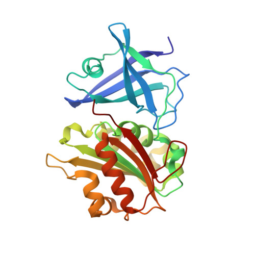
2QDX - PubMed Abstract:
Among the 118 genes upregulated by Pseudomonas aeruginosa in response to iron starvation [Ochsner, U. A., Wilderman, P. J., Vasil, A. I., and Vasil, M. L. (2002) Mol. Microbiol. 45, 1277-1287], we focused on the products of the two genes encoding electron transfer proteins, as a means of identifying the redox partners of the heme oxygenase (pa-HO) expressed under low-iron stress conditions. Biochemical and spectroscopic investigations demonstrated that the bfd gene encodes a 73-amino acid protein (pa-Bfd) that incorporates a [2Fe-2S]2+/+ center, whereas the fpr gene encodes a 258-residue NADPH-dependent ferredoxin reductase (pa-FPR) that utilizes FAD as a cofactor. In vitro reconstitution of pa-HO catalytic activity with the newly characterized proteins led to the surprising observation that pa-FPR efficiently supports the catalytic cycle of pa-HO, without the need of a ferredoxin. In comparison, electron transfer from pa-Bfd to pa-HO is sluggish, which strongly argues against the possibility that the seven electrons needed by pa-HO to degrade biliverdin are transferred from NADPH to pa-HO in a ferredoxin (Bfd)-dependent manner. Given that pa-HO functions to release iron from exogenous heme acquired under iron-starvation conditions, the use of a flavoenzyme rather than an iron-sulfur center-containing protein to support heme degradation is an efficient use of resources in the cell. The crystal structure of pa-FPR (1.6 A resolution) showed that its fold is comparable that of the superfamily of ferredoxin reductases and most similar to the structure of Azotobacter vinelandii FPR and Escherichia coli flavodoxin reductase. The latter two enzymes interact with distinct redox partners, a ferredoxin and a flavodoxin, respectively. Hence, findings reported herein extend the range of redox partners recognized by the fold of pa-FPR to include a heme oxygenase (pa-HO).
Organizational Affiliation:
Ralph N. Adams Institute for Bioanalytical Chemistry and Department of Chemistry, University of Kansas, Multidisciplinary Research Building, 2030 Becker Drive, Room 220 E, Lawrence, Kansas 66047, USA.