2Q8K

The crystal structure of Ebp1


Experimental Data Snapshot

  • Method: X-RAY DIFFRACTION
  • Resolution: 1.60 Å
  • R-Value Free: 0.219 
  • R-Value Work: 0.182 
  • R-Value Observed: 0.184 

wwPDB Validation   3D Report Full Report


This is version 1.2 of the entry. See complete history


Literature

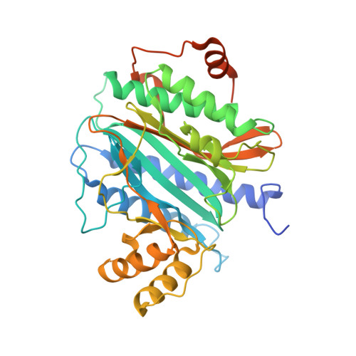
The crystal structure of Ebp1 reveals a methionine aminopeptidase fold as binding platform for multiple interactions.

Kowalinski, E.Bange, G.Bradatsch, B.Hurt, E.Wild, K.Sinning, I.

(2007) FEBS Lett 581: 4450-4454

  • DOI: https://doi.org/10.1016/j.febslet.2007.08.024
  • Primary Citation of Related Structures:  
    2Q8K

  • PubMed Abstract: 

    The ErbB-3 receptor binding protein (Ebp1) is a member of the proliferation-associated 2G4 (PA2G4) family implicated in regulation of cell growth and differentiation. Here, we report the crystal structure of the human Ebp1 at 1.6 A resolution. The protein has the conserved pita bread fold of methionine aminopeptidases, but without the characteristic enzymatic activity. Moreover, Ebp1 is known to interact with a number of proteins and RNAs involved in either transcription regulation or translation control. The structure provides insights in how Ebp1 discriminates between its different interaction partners.


  • Organizational Affiliation

    Heidelberg University Biochemistry Center (BZH), INF 328, D-69120 Heidelberg, Germany.


Macromolecules
Find similar proteins by:  (by identity cutoff)  |  3D Structure
Entity ID: 1
MoleculeChains Sequence LengthOrganismDetailsImage
Proliferation-associated protein 2G4401Homo sapiensMutation(s): 0 
Gene Names: PA2G4EBP1
UniProt & NIH Common Fund Data Resources
Find proteins for Q9UQ80 (Homo sapiens)
Explore Q9UQ80 
Go to UniProtKB:  Q9UQ80
PHAROS:  Q9UQ80
GTEx:  ENSG00000170515 
Entity Groups  
Sequence Clusters30% Identity50% Identity70% Identity90% Identity95% Identity100% Identity
UniProt GroupQ9UQ80
Sequence Annotations
Expand
  • Reference Sequence
Experimental Data & Validation

Experimental Data

  • Method: X-RAY DIFFRACTION
  • Resolution: 1.60 Å
  • R-Value Free: 0.219 
  • R-Value Work: 0.182 
  • R-Value Observed: 0.184 
  • Space Group: P 41 21 2
Unit Cell:
Length ( Å )Angle ( ˚ )
a = 73.887α = 90
b = 73.887β = 90
c = 183.348γ = 90
Software Package:
Software NamePurpose
REFMACrefinement
ADSCdata collection
MOSFLMdata reduction
SCALAdata scaling
PHASERphasing

Structure Validation

View Full Validation Report


Entry History 

Deposition Data

Revision History  (Full details and data files)

  • Version 1.0: 2007-09-25
    Type: Initial release
  • Version 1.1: 2011-07-13
    Changes: Non-polymer description, Version format compliance
  • Version 1.2: 2023-08-30
    Changes: Data collection, Database references, Derived calculations, Refinement description