2Q3X

The RIM1alpha C2B domain


Experimental Data Snapshot

  • Method: X-RAY DIFFRACTION
  • Resolution: 1.73 Å
  • R-Value Free: 0.215 
  • R-Value Work: 0.178 
  • R-Value Observed: 0.179 

wwPDB Validation   3D Report Full Report


This is version 1.1 of the entry. See complete history


Literature

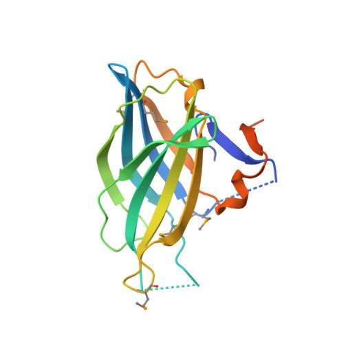
Crystal Structure of the RIM1alpha C(2)B Domain at 1.7 A Resolution.

Guan, R.Dai, H.Tomchick, D.R.Dulubova, I.Machius, M.Sudhof, T.C.Rizo, J.

(2007) Biochemistry 46: 8988-8998

  • DOI: https://doi.org/10.1021/bi700698a
  • Primary Citation of Related Structures:  
    2Q3X

  • PubMed Abstract: 

    RIM proteins play critical roles in synaptic vesicle priming and diverse forms of presynaptic plasticity. The C-terminal C2B domain is the only module that is common to all RIMs but is only distantly related to well-studied C2 domains, and its three-dimensional structure and interactions have not been characterized in detail. Using NMR spectroscopy, we now show that N- and C-terminal extensions beyond the predicted C2B domain core sequence are necessary to form a folded, stable RIM1alpha C2B domain. We also find that the isolated RIM1alpha C2B domain is not sufficient for previously described protein-protein interactions involving the RIM1alpha C-terminus, suggesting that additional sequences adjacent to the C2B domain might be required for these interactions. However, analytical ultracentrifugation shows that the RIM1alpha C2B domain forms weak dimers in solution. The crystal structure of the RIM1alpha C2B domain dimer at 1.7 A resolution reveals that it forms a beta-sandwich characteristic of C2 domains and that the unique N- and C-terminal extensions form a small subdomain that packs against the beta-sandwich and mediates dimerization. Our results provide a structural basis to understand the function of RIM C2B domains and suggest that dimerization may be a crucial aspect of RIM function.


  • Organizational Affiliation

    Department of Biochemistry, University of Texas Southwestern Medical Center, 6000 Harry Hines Boulevard, Dallas, Texas 75390, USA.


Macromolecules
Find similar proteins by:  (by identity cutoff)  |  3D Structure
Entity ID: 1
MoleculeChains Sequence LengthOrganismDetailsImage
Regulating synaptic membrane exocytosis protein 1
A, B
171Rattus norvegicusMutation(s): 5 
Gene Names: Rims1Rim1
UniProt
Find proteins for Q9JIR4 (Rattus norvegicus)
Explore Q9JIR4 
Go to UniProtKB:  Q9JIR4
Entity Groups  
Sequence Clusters30% Identity50% Identity70% Identity90% Identity95% Identity100% Identity
UniProt GroupQ9JIR4
Sequence Annotations
Expand
  • Reference Sequence
Small Molecules
Ligands 3 Unique
IDChains Name / Formula / InChI Key2D Diagram3D Interactions
SO4
Query on SO4

Download Ideal Coordinates CCD File 
C [auth A]
D [auth A]
E [auth A]
F [auth A]
G [auth A]
C [auth A],
D [auth A],
E [auth A],
F [auth A],
G [auth A],
J [auth B],
K [auth B]
SULFATE ION
O4 S
QAOWNCQODCNURD-UHFFFAOYSA-L
CL
Query on CL

Download Ideal Coordinates CCD File 
H [auth A],
I [auth A],
M [auth B]
CHLORIDE ION
Cl
VEXZGXHMUGYJMC-UHFFFAOYSA-M
NA
Query on NA

Download Ideal Coordinates CCD File 
L [auth B]SODIUM ION
Na
FKNQFGJONOIPTF-UHFFFAOYSA-N
Modified Residues  1 Unique
IDChains TypeFormula2D DiagramParent
MSE
Query on MSE
A, B
L-PEPTIDE LINKINGC5 H11 N O2 SeMET
Experimental Data & Validation

Experimental Data

  • Method: X-RAY DIFFRACTION
  • Resolution: 1.73 Å
  • R-Value Free: 0.215 
  • R-Value Work: 0.178 
  • R-Value Observed: 0.179 
  • Space Group: P 31 2 1
Unit Cell:
Length ( Å )Angle ( ˚ )
a = 62.037α = 90
b = 62.037β = 90
c = 145.18γ = 120
Software Package:
Software NamePurpose
REFMACrefinement
ADSCdata collection
HKL-2000data reduction
HKL-2000data scaling
SHELXSphasing

Structure Validation

View Full Validation Report


Entry History 

Deposition Data

Revision History  (Full details and data files)

  • Version 1.0: 2007-08-28
    Type: Initial release
  • Version 1.1: 2011-07-13
    Changes: Advisory, Derived calculations, Version format compliance